Review
. 2021 Apr 1;34(2):246-257.
doi: 10.1097/WCO.0000000000000913.

Cerebral small vessel disease and vascular cognitive impairment: from diagnosis to management

Affiliations
Review

Cerebral small vessel disease and vascular cognitive impairment: from diagnosis to management

Maria Clara Zanon Zotin et al. Curr Opin Neurol. .

Abstract

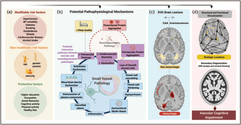
Purpose of review: We present recent developments in the field of small vessel disease (SVD)-related vascular cognitive impairment, including pathological mechanisms, updated diagnostic criteria, cognitive profile, neuroimaging markers and risk factors. We further address available management and therapeutic strategies.

Recent findings: Vascular and neurodegenerative pathologies often co-occur and share similar risk factors. The updated consensus criteria aim to standardize vascular cognitive impairment (VCI) diagnosis, relying strongly on cognitive profile and MRI findings. Aggressive blood pressure control and multidomain lifestyle interventions are associated with decreased risk of cognitive impairment, but disease-modifying treatments are still lacking. Recent research has led to a better understanding of mechanisms leading to SVD-related cognitive decline, such as blood-brain barrier dysfunction, reduced cerebrovascular reactivity and impaired perivascular clearance.

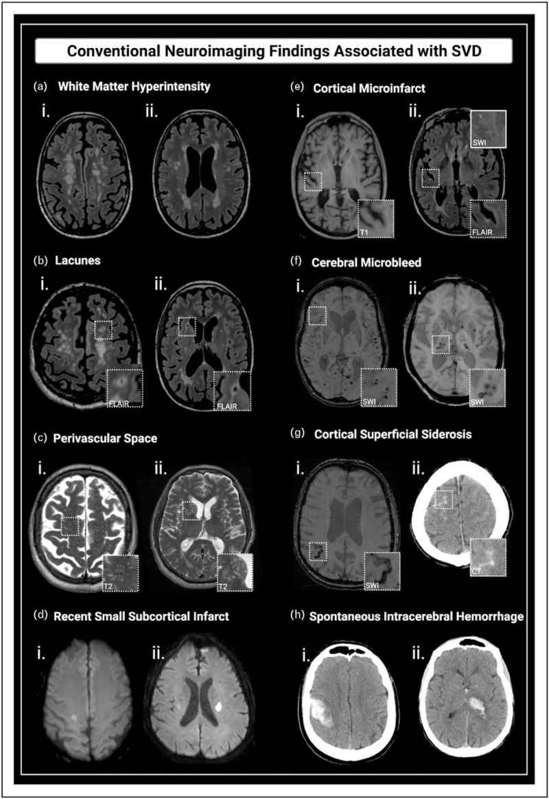
Summary: SVD is the leading cause of VCI and is associated with substantial morbidity. Tackling cardiovascular risk factors is currently the most effective approach to prevent cognitive decline in the elderly. Advanced imaging techniques provide tools for early diagnosis and may play an important role as surrogate markers for cognitive endpoints in clinical trials. Designing and testing disease-modifying interventions for VCI remains a key priority in healthcare.


Conflict of interest statement

There are no conflicts of interest.

Figures

Box 1
Box 1
no caption available
FIGURE 1
FIGURE 1
Schematic overview of potential mechanisms leading to vascular cognitive impairment. (a) Risk factors associated with SVD and related cognitive decline. (b) Potential pathophysiological mechanisms of SVD. Dysfunctional NVUs have an important role in early SVD pathology. Several effects are described around the blue circle, the order of which is not yet established. Combined, these effects are thought to contribute to exacerbate tissue injury. (c) Typical brain lesions associated with sporadic SVD: CAA (left) and arteriolosclerosis (right) patterns. The hemorrhagic lesions (bottom figure) include: CMB, cSS, SAH and ICH. The non-hemorrhagic lesions (upper figure) include WMH, lacunes, PVS, small acute subcortical infarcts and cortical CMI. (d) Potential mechanisms involved in SVD-related cognitive decline: impairment of structural and functional connectivity (upper figure) and secondary degeneration (lower figure). AD, Alzheimer's disease; CAA, cerebral amyloid angiopathy; CMB, cerebral microbleed; CMI, cerebral microinfarcts; cSS, cortical superficial siderosis; ICH, intracerebral hemorrhage; NVU, neurovascular unit; PVS, perivascular spaces; SAH, subarachnoid hemorrhage; SVD, small vessel disease; WMH, white matter hyperintensity. Adapted from [,▪▪,14,15]. Created with BioRender.com.
FIGURE 2
FIGURE 2
Conventional neuroimaging findings associated with SVD. (a) WMH: confluent hyperintensity foci visible on FLAIR (i and ii). (b) Lacunes: fluid-filled subcortical cavities, 3–15 mm, isointense to CSF, often with hyperintense rims on FLAIR (i - lobar lacune; ii - deep lacune). (c) PVS: linear, ovoid or round-shaped fluid-filled spaces, following the course of vessels (i - predominating in the centrum semiovale; ii - affecting the basal ganglia). (d) Recent small subcortical infarcts: hyperintense foci on DWI (i and ii). (e) Cortical CMIs: intracortical lesions ≤ 4 mm, hypointense on T1 (i) and hyper or isointense on FLAIR (ii). (f) CMBs: foci of hemosiderin deposition, with very low signal intensity on SWI (lobar CMBs (i), and deep CMBs (ii)). (g) cSS: linear hypointense foci with gyriform pattern over the cerebral cortex on SWI (i). The acute form of superficial bleeding is cSAH, seen as linear hyperdensities on CT (ii) or as hyperintensity on FLAIR. (h) Spontaneous ICH: nontraumatic lobar (i) and deep (ii) hemorrhages, depicted as focal hyperdense lesions on CT. CAA, cerebral amyloid angiopathy; CMB, cerebral microbleed; CMI, cerebral microinfarcts; cSAH, convexity subarachnoid hemorrhage; CSF, cerebrospinal fluid; cSS, cortical superficial siderosis; CT, computed tomography; DWI, diffusion weighted image; FLAIR, Fluid-attenuated inversion recovery; ICH, intracerebral hemorrhage; PVS, perivascular space; SVD, small vessel disease; SWI, susceptibility weighted imaging; WMH, white matter hyperintensity. Adapted from [14]. Created with BioRender.com.

Similar articles

Cited by

References

    1. Flier WM, van der, Skoog I, et al. . Vascular cognitive impairment. Nat Rev Dis Primers 2018; 4:18003. - PubMed
    1. Wolters FJ, Ikram MA. Epidemiology of vascular dementia. Arterioscler Thromb Vasc Biol 2019; 39:1542–1549. - PubMed
    2. This review highlights the multifactorial nature of cognitive decline in the elderly, and suggests that definitions and classifications in the field of dementia should acknowledge the potential contributions from different pathways.

    1. Boyle PA, Yu L, Wilson RS, et al. . Person-specific contribution of neuropathologies to cognitive loss in old age. Ann Neurol 2018; 83:74–83. - PMC - PubMed
    2. A recent clinical–pathologic populational study on the prevalence and co-occurrence of multiple neuropathologies in the elderly, quantifying their contributions to cognitive loss at the individual level.

    1. Viswanathan A, Rocca WA, Tzourio C. Vascular risk factors and dementia. Neurology 2009; 72:368–374. - PMC - PubMed
    1. Dichgans M, Leys D. Vascular cognitive impairment. Circ Res 2017; 120:573–591. - PubMed

Publication types

MeSH terms