. 2021 Feb 25;17(2):e1009355.
doi: 10.1371/journal.ppat.1009355. eCollection 2021 Feb.

An aspartyl protease-mediated cleavage regulates structure and function of a flavodoxin-like protein and aids oxidative stress survival

Affiliations

An aspartyl protease-mediated cleavage regulates structure and function of a flavodoxin-like protein and aids oxidative stress survival

Anamika Battu et al. PLoS Pathog. .

Abstract

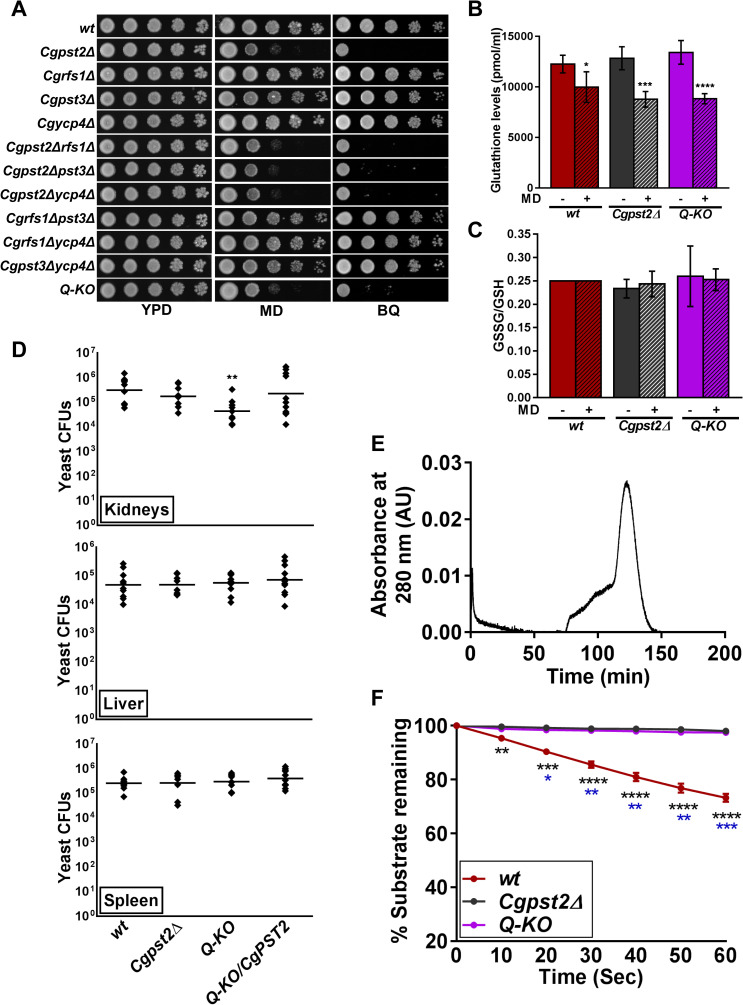
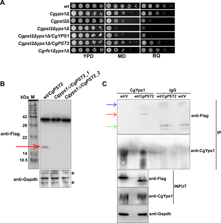
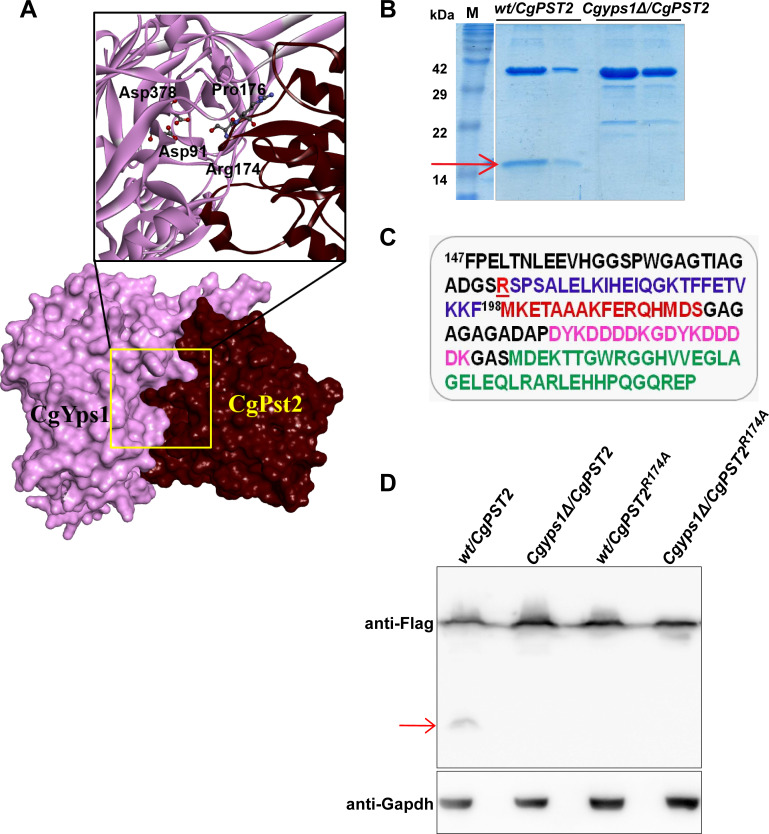
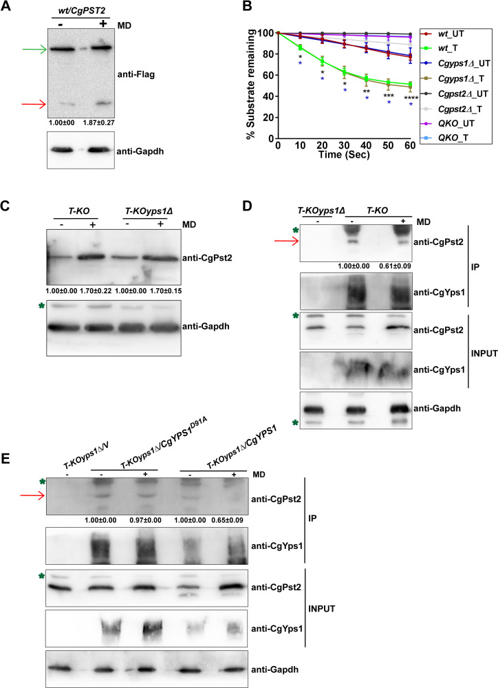
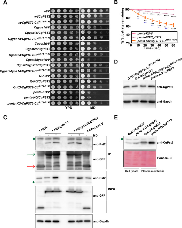
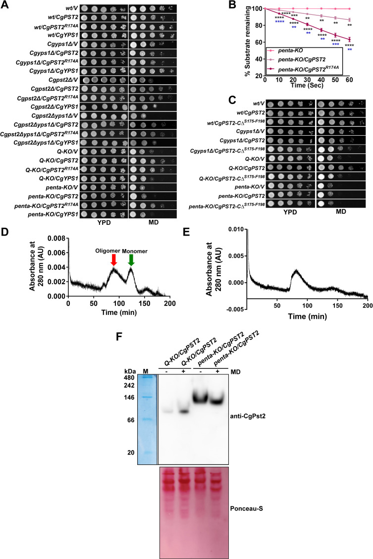
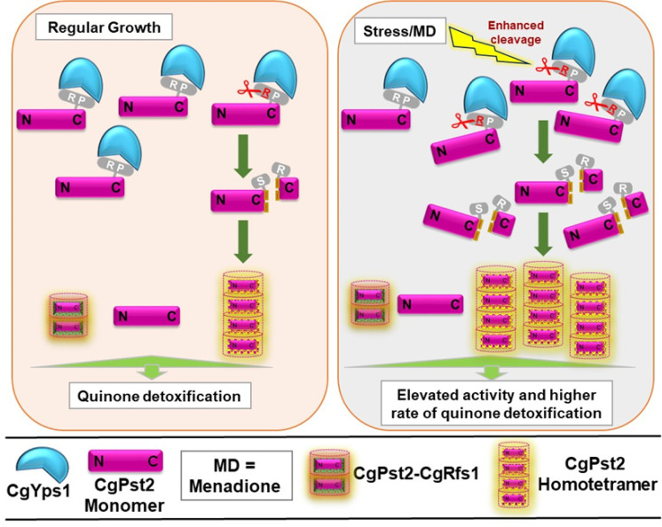
A family of eleven glycosylphosphatidylinositol-anchored aspartyl proteases, commonly referred to as CgYapsins, regulate a myriad of cellular processes in the pathogenic yeast Candida glabrata, but their protein targets are largely unknown. Here, using the immunoprecipitation-mass spectrometry approach, we identify the flavodoxin-like protein (Fld-LP), CgPst2, to be an interactor of one of the aspartyl protease CgYps1. We also report the presence of four Fld-LPs in C. glabrata, which are required for survival in kidneys in the murine model of systemic candidiasis. We further demonstrated that of four Fld-LPs, CgPst2 was solely required for menadione detoxification. CgPst2 was found to form homo-oligomers, and contribute to cellular NADH:quinone oxidoreductase activity. CgYps1 cleaved CgPst2 at the C-terminus, and this cleavage was pivotal to oligomerization, activity and function of CgPst2. The arginine-174 residue in CgPst2 was essential for CgYps1-mediated cleavage, with alanine substitution of the arginine-174 residue also leading to elevated activity and oligomerization of CgPst2. Finally, we demonstrate that menadione treatment led to increased CgPst2 and CgYps1 protein levels, diminished CgYps1-CgPst2 interaction, and enhanced CgPst2 cleavage and activity, thereby implicating CgYps1 in activating CgPst2. Altogether, our findings of proteolytic cleavage as a key regulatory determinant of CgPst2, which belongs to the family of highly conserved, electron-carrier flavodoxin-fold-containing proteins, constituting cellular oxidative stress defense system in diverse organisms, unveil a hidden regulatory layer of environmental stress response mechanisms.


Conflict of interest statement

The authors have declared that no competing interests exist.

Figures

Fig 1
Fig 1. The flavodoxin-like protein CgPst2 plays an essential role in menadione and benzoquinone resistance.
A. Serial dilution spotting assay illustrating MD (95 μM) and BQ (4 mM) sensitivity of indicated C. glabrata mutants. The Q-KO strain lacks four flavodoxin-like proteins, CgPst2, CgRfs1, CgPst3 and CgYcp4. B. Total intracellular glutathione content of log-phase cells (10.0 OD600). Data (mean ± SEM; n = 4–5) represent total glutathione concentration normalized to one ml of cell lysate. *, p ≤ 0.05, ***, p ≤ 0.001, ****, p ≤ 0.0001; unpaired two-tailed Student’s t test. C. Ratio of oxidized (GSSG) to reduced (GSH) glutathione. 4-vinylpyridine was used to derivatize/block all free thiols in cell lysates, followed by GSSG estimation using DTNB [5,5-dithio-bis-(2-nitrobenzoic acid); Ellman’s Reagent]. Reduced GSH levels were calculated by subtracting GSSG levels from total glutathione levels. Data represent (mean ± SEM; n = 3–5). D. Survival of indicated C. glabrata strains in the murine model of systemic candidiasis. Six to eight-week-old female BALB/c mice were infected with cells (4X10⁷ cells; 100 μl cell suspension) of overnight YPD/CAA-grown C. glabrata strains, wt, Cgpst2Δ, Q-KO and Q-KO expressing CgPST2, through tail vein injections. At 7th day post infection, mice were sacrificed, target organs (kidneys, liver and spleen) collected, homogenized in sterile PBS and appropriate homogenate dilutions were plated on YPD medium containing penicillin and streptomycin antibiotics. Total fungal burden in each mouse organ was determined, and plotted. Diamonds and bars indicate CFUs recovered from an individual mouse, and CFU geometric mean (n = 8–12), respectively, for each organ. **, p < 0.01; Mann-Whitney U-test. E. Size exclusion chromatogram of purified CgPst2 protein. 300 μg 6XHIS-FLAG-CgPst2 was applied to a Sephacryl S-200 column, and protein elution profiles were determined using the absorbance values at 280 nm. The peak corresponds to 29 kDa CgPst2 protein. AU, Arbitrary Units. F. NADH:quinone oxidoreductase activity in cell lysates (500 μg) of log-phase wt, Cgpst2Δ and Q-KO cells, as measured using menadione (500 μM) and NADH (500 μM) substrates. Absorbance of the substrate NADH was considered as 100 at 0 h time point, and NADH oxidation was deduced from the formula: [(absorbance at each time point/0 h absorbance) X 100]. Data represent mean ± SEM. Black and blue asterisks indicate statistically significant differences in activity between wt and Cgpst2Δ, and wt and Q-KO strains, respectively. *, p < 0.0332; **, p < 0.0021; ***, p < 0.0002; ****, p < 0.0001; Grouped multiple t-test (n = 3 to 4).
Fig 2
Fig 2. CgPst2 interacts with CgYps1.
A. Serial dilution spotting assay illustrating MD (95 μM) and BQ (4 mM) sensitivity of indicated CgYPS1-deleted strains. B. Immunoblot analysis of CgPst2-SFB in cell extracts (60 μg) of wt and Cgyps1Δ transformants expressing CgPST2-SFB. The red arrow marks the small (~ 16 kDa) cleaved fragment of CgPst2. Cgyps1Δ/CgPST2_1 and Cgyps1Δ/CgPST2_2 represent two independent Cgyps1Δ transformants. M, Protein Marker. C. Immunoblot analysis of CgPst2-CgYps1 interaction. Lysates (3.0 mg) of wt cells expressing either SFB-GFP or CgPst2-SFB were immunoprecipitated with anti-CgYps1 or anti-mouse immunoglobulin G (IgG) antibody, resolved on 4–20% polyacrylamide gradient, 12% polyacrylamide and 12% polyacrylamide gels for CgYps1, CgPst2-SFB and CgGapdh, respectively, and were probed with anti-CgYps1, anti-Flag and anti-Gapdh antibodies. CgGapdh was used as loading control. Please note that for Input samples, 200, 75 and 75 μg protein was loaded for detection of CgYps1, CgPst2-SFB and CgGapdh proteins, respectively. Red, blue and green arrows mark the CgPst2-SFB, Ig heavy chain and Ig light chain, respectively. ‘V’ refers to the vector expressing SFB-GFP protein.
Fig 3
Fig 3. CgPst2 is cleaved at the C-terminus.
A. Molecular surface and Ribbon models depicting CgYps1 (Length: 601 aa; Predicted MW: 64 kDa) with CgPst2 (Length: 198 aa; Predicted MW: 21 kDa). D91 and D378 in CgYps1 represent catalytically active aspartic acid residues. The 171DGSRSPSA178 region in CgPst2 contains predicted CgYps1-binding residues, Arginine-174 and Proline-176. Hydrogen bond-forming amino acid residues are highlighted in bold letters in the predicted interaction region. The molecular surfaces of receptor (CgYps1) and substrate (CgPst2) are coloured purple and maroon, respectively. B. Coomassie-blue-stained 18% SDS-PAGE gel images indicating expression of the full-length (~ 40 kDa) and the C-terminal cleaved fragment (~ 16 kDa; marked with a red arrow) of CgPst2-SFB, after two-step affinity purification of cell extracts of wt and Cgyps1Δ strains. C. The C-terminus amino acid sequence of CgPst2-SFB protein starting with the phenylalanine (F) residue at 147th position of CgPst2 protein. CgPst2 is 198 amino acid long. The predicted cleavage residue R174 is underlined and indicated in red. The SFB tag (85 aa) consists of S-protein (MKETAAAKFERQHMDS; brown), two copies of the FLAG tag (DYKDDDDK; pink) and streptavidin-binding-peptide (MDEKTTGWRGGHVVEGLAGELEQLRARLEHHPQGQREP; green) sequences. MS analysis of the shorter CgPst2-SFB fragment identified peptides corresponding to SFB and CgPst2 sequences. D. Immunoblot analysis of wt cell extracts expressing either wild-type CgPst2-SFB or alanine-substituted-CgPst2-SFB (CgPST2R174A), using anti-Flag antibody. The red arrow marks the small cleaved fragment of wild-type CgPst2. CgGapdh was used as a loading control.
Fig 4
Fig 4. The cleavage and NADH:quinone oxidoreductase activity of CgPst2 is increased upon menadione treatment.
A. Immunoblot analysis of CgPst2 expression, using anti-Flag antibody, in cell extracts of untreated or menadione-(MD; 90 μM for 90 min)-treated log-phase cells of the wt strain expressing CgPst2-SFB, using anti-Flag antibody. CgGapdh was used as a loading control. The intensity of the shorter 16 kDa band in 4 independent Western blots was quantified using the ImageJ densitometry software, and this signal was normalized to the corresponding CgGapdh-normalized total CgPst2 signal, as total CgPst2 levels were also elevated, upon MD treatment, compared to CgGapdh signal. Data (mean ± SEM) represent the fold-change in levels of the C-terminal cleaved fragment of CgPst2-SFB in treated cells, compared to untreated cells (considered as 1.0), and are shown underneath the blot. p ≤ 0.05; paired two-tailed Student’s t test. The red and green arrows indicate the C-terminal cleaved fragment of CgPst2 and full length CgPst2-SFB, respectively. B. NADH:quinone oxidoreductase activity in log-phase cultures of indicated C. glabrata strains that were either treated with 90 μM menadione (T) for 90 min, or left untreated (UT), as measured using menadione (500 μM) and NADH (500 μM) substrates. Absorbance of the substrate NADH was considered as 100 at 0 h time point, and NADH oxidation was deduced from the formula: [(absorbance at each time point/0 h absorbance) X 100]. Black and blue asterisks indicate statistically significant differences in activity of treated wt and Cgyps1Δ samples, respectively, compared to respective untreated lysates. The Q-KO strain lacks four flavodoxin-like proteins, CgPst2, CgRfs1, CgPst3 and CgYcp4. *, p < 0.0332; **, p < 0.0021; ***, p < 0.0002; ****, p < 0.0001; Grouped multiple t-test (n = 3 to 5). C. Immunoblot analysis of CgPst2 levels in T-KO (Cgrfs1Δpst3Δycp4Δ) and T-KOyps1Δ (Cgrfs1Δpst3Δycp4Δyps1Δ) cells, using anti-CgPst2 antibody. The intensity of individual bands in 4 independent Western blots was quantified using ImageJ densitometry software, and CgPst2 signal was normalized to the corresponding CgGapdh signal. Fold-change (mean ± SEM) in CgPst2 levels in treated cells, compared to untreated cells (considered as 1.0), is shown underneath the blot. p ≤ 0.05; paired two-tailed Student’s t test. The green asterisk indicates non-specific band. D-E. Immunoblot analysis showing CgYps1-CgPst2 (D) and CgYps1-CgYps1D91A-CgPst2 (E) interaction upon MD treatment (90 μM for 90 min) in T-KO (Cgrfs1Δpst3Δycp4Δ) and T-KOyps1Δ (Cgrfs1Δpst3Δycp4Δyps1Δ) cells, using anti-CgPst2 antibody. 6 mg precleared cell lysates were incubated with anti-CgYps1 antibody-conjugated beads for 12 h at 4°C. After bead washing, bead-bound proteins were boiled in 2X-SDS loading buffer and resolved on 4–20% polyacrylamide gradient and 12% polyacrylamide gels for CgYps1 and CgPst2 samples, respectively. For input samples, 60, 60 and 200 μg protein were loaded to detect CgPst2, CgGapdh and CgYps1 proteins, respectively. Two to three independent Western blots were quantified using the ImageJ densitometry software, and CgPst2 signal was normalized to the corresponding CgGapdh signal. Fold-change in CgPst2 levels in treated cells, compared to untreated cells (considered as 1.0), is shown underneath the blot. p ≤ 0.05; paired two-tailed Student’s t test. The red arrow marks CgPst2 band, while the green asterisk denotes non-specific band.
Fig 5
Fig 5. CgPst2-CΔR174-F198 possesses higher activity than CgPst2 in penta-KO.
A. Serial dilution spotting assay showing CgPST2-CΔR174-F198-mediated rescue of MD (80 μM) sensitivity. V, pRK74 vector. The Q-KO strain lacks four flavodoxin-like proteins (Fld-LPs), CgPst2, CgRfs1, CgPst3 and CgYcp4, while the penta-KO strain lacks CgYps1 along with four Fld-LPs, CgPst2, CgRfs1, CgPst3 and CgYcp4. B. NADH:quinone oxidoreductase activity in cell extracts of penta-KO expressing CgPST2 or CgPST2-CΔR174-F198. Data represent mean ± SEM (n = 3). Black and blue asterisks represent statistically significant activity differences in indicated strains compared to penta-KO/V and penta-KO/CgPST2, respectively. *, p < 0.0332; **, p < 0.0021; ***, p < 0.0002; ****, p < 0.0001; Grouped multiple t-test. V, pRK74 vector. C. Immunoblot analysis showing interaction between CgPst2 and CgRfs1-GFP. 3 mg precleared lysates of untreated and MD (90 μM for 90 min)-treated T-KO (Cgrfs1Δpst3Δycp4Δ) and T-KOyps1Δ (Cgrfs1Δpst3Δycp4Δyps1Δ) cells expressing CgRfs1-GFP were incubated with anti-GFP antibody-conjugated beads, followed by Western analysis. The red and green arrows mark GFP and CgRfs1-GFP protein bands, respectively, in IP samples, while the green asterisk denotes non-specific band. D. Immunoblot analysis of CgPst2 levels in Q-KO and penta-KO strains expressing CgPST2 or CgPST2-CΔR174-F198. E. Immunoblot analysis showing enrichment of CgPst2 in plasma membrane fractions of the Q-KO and penta-KO strains expressing CgPST2. Whole-cell lysates and plasma membrane fractions (60 μg), prepared by glass bead lysis and sucrose gradient ultracentrifugation, respectively, were resolved on 12% polyacrylamide gel, and probed with anti-CgPst2 antibody. The ponceau S-stained membrane is shown as loading control. The green asterisk denotes non-specific band.
Fig 6
Fig 6. CgPst2R174A rescues MD sensitivity of CgPST2-deleted strains.
A. Serial dilution spotting assay showing CgPST2R174A-mediated rescue of MD (80 μM) sensitivity. V, pRK74 vector. The Q-KO strain lacks four flavodoxin-like proteins (Fld-LPs), CgPst2, CgRfs1, CgPst3 and CgYcp4, while the penta-KO strain lacks CgYps1 along with four Fld-LPs, CgPst2, CgRfs1, CgPst3 and CgYcp4. B. NADH:quinone oxidoreductase activity in cell extracts of penta-KO expressing CgPST2 or CgPST2R174A. Data represent mean ± SEM (n = 3). Black and blue asterisks represent statistically significant activity differences in indicated strains compared to penta-KO and penta-KO/CgPST2, respectively. **, p < 0.0021; ***, p < 0.0002; ****, p < 0.0001; Grouped multiple t-test. C. Serial dilution spotting assay showing MD (80 μM) sensitivity of Q-KO and penta-KO strains expressing CgPST2-CΔS175-F198. D-E. Size exclusion chromatograms of purified CgPst2R174A (D) and CgPst2-CΔR174-F198 (E). After loading on the Sephacryl S-200 column, protein elution profiles were determined using the absorbance values at 280 nm. The CgPst2R174A showed two peaks corresponding to 296 kDa (red arrow; oligomeric form) and 29 kDa (green arrow; monomeric form) sizes, and CgPst2-CΔR174-F198 showed one peak of 296 kDa. F. Native PAGE analysis showing increased oligomer formation upon MD treatment. 200 μg whole cell lysates of untreated and MD (90 μM for 90 min)-treated Q-KO and penta-KO expressing CgPST2 were resolved in a discontinuous Tris-glycine buffer system under non-denaturing conditions, and probed with anti-CgPst2 antibody. The native protein molecular weight marker (M) was stained with coomassie brilliant blue, and is shown on the right side of the blot. The ponceau S-stained membrane is shown as loading control.
Fig 7
Fig 7. A schematic summarizing key findings of the study.
Arginine-174 (R) and Proline-176 (P) residues of CgPst2 are predicted to interact with CgYps1 at the plasma membrane, and CgYps1 processes R174 residue in the C-terminus of CgPst2. This cleavage, which is elevated upon menadione treatment, leads to removal of the C-terminal domain, resulting in CgPst2 homo-tetramerization, higher activity and efficient quinone detoxification. Of note, the signal, that stimulates CgYps1-mediated cleavage of CgPst2, is not known. CgPst2 also interacts with CgRfs1, however, CgPst2-CgRfs1 association is not dependent on CgYps1, and occurs under both regular and MD treatment conditions. Altogether, CgPst2 functions are regulated at multiple levels, and CgYps1-mediated cleavage of CgPst2 may reflect one of many mechanisms controlling CgPst2 activity.

References

    1. Brown GD, Denning DW, Gow NAR, Levitz SM, Netea MG, White TC. Hidden killers: human fungal infections. Sci Transl Med. 2012;4: 165rv13. 10.1126/scitranslmed.3004404 - DOI - PubMed
    1. Bassetti M, Merelli M, Righi E, Diaz-Martin A, Rosello EM, Luzzati R, et al. Epidemiology, Species Distribution, Antifungal Susceptibility, and outcome of Candidemia across five sites in Italy and Spain. J Clin Microbiol. 2013;51: 4167–4172. 10.1128/JCM.01998-13 - DOI - PMC - PubMed
    1. Lamoth F, Lockhart SR, Berkow EL, Calandra T. Changes in the epidemiological landscape of invasive candidiasis. J Antimicrob Chemother. 2018;73: i4–i13. 10.1093/jac/dkx444 - DOI - PMC - PubMed
    1. Pappas PG, Lionakis MS, Arendrup MC, Ostrosky-Zeichner L, Kullberg BJ. Invasive candidiasis. Nat Rev Dis Prim. 2018;4: 1–20. 10.1038/s41572-018-0001-z - DOI - PubMed
    1. Pfaller MA, Diekema DJ, Turnidge JD, Castanheira M, Jones RN. Twenty years of the SENTRY antifungal surveillance program: Results for Candida species from 1997–2016. Open forum Infect Dis. 2019;6: S79–S94. 10.1093/ofid/ofy358 - DOI - PMC - PubMed

Publication types

MeSH terms