

Bacterial virulence plays a crucial role in MRSA sepsis
- PMID: 33630954
- PMCID: PMC7942999
- DOI: 10.1371/journal.ppat.1009369
Bacterial virulence plays a crucial role in MRSA sepsis
Abstract
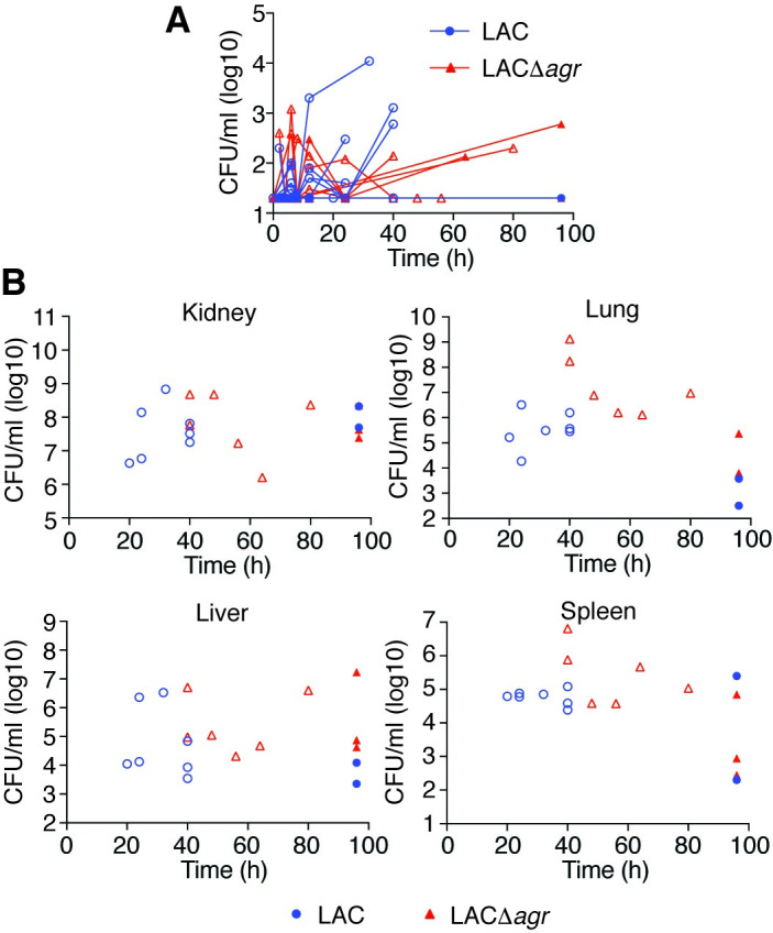
Bacterial sepsis is a major global cause of death. However, the pathophysiology of sepsis has remained poorly understood. In industrialized nations, Staphylococcus aureus represents the pathogen most commonly associated with mortality due to sepsis. Because of the alarming spread of antibiotic resistance, anti-virulence strategies are often proposed to treat staphylococcal sepsis. However, we do not yet completely understand if and how bacterial virulence contributes to sepsis, which is vital for a thorough assessment of such strategies. We here examined the role of virulence and quorum-sensing regulation in mouse and rabbit models of sepsis caused by methicillin-resistant S. aureus (MRSA). We determined that leukopenia was a predictor of disease outcome during an early critical stage of sepsis. Furthermore, in device-associated infection as the most frequent type of staphylococcal blood infection, quorum-sensing deficiency resulted in significantly higher mortality. Our findings give important guidance regarding anti-virulence drug development strategies for the treatment of staphylococcal sepsis. Moreover, they considerably add to our understanding of how bacterial sepsis develops by revealing a critical early stage of infection during which the battle between bacteria and leukocytes determines sepsis outcome. While sepsis has traditionally been attributed mainly to host factors, our study highlights a key role of the invading pathogen and its virulence mechanisms.
Conflict of interest statement
The authors have declared that no competing interests exist.
Figures


Similar articles
-
Quorum Sensing, Virulence, and Antibiotic Resistance of USA100 Methicillin-Resistant Staphylococcus aureus Isolates.mSphere. 2019 Aug 14;4(4):e00553-19. doi: 10.1128/mSphere.00553-19. mSphere. 2019. PMID: 31413175 Free PMC article.
-
Pathogenicity and virulence of Staphylococcus aureus.Virulence. 2021 Dec;12(1):547-569. doi: 10.1080/21505594.2021.1878688. Virulence. 2021. PMID: 33522395 Free PMC article. Review.
-
Redeploying β-Lactam Antibiotics as a Novel Antivirulence Strategy for the Treatment of Methicillin-Resistant Staphylococcus aureus Infections.J Infect Dis. 2017 Jan 1;215(1):80-87. doi: 10.1093/infdis/jiw461. Epub 2016 Nov 14. J Infect Dis. 2017. PMID: 28077586 Free PMC article.
-
VraSR and Virulence Trait Modulation during Daptomycin Resistance in Methicillin-Resistant Staphylococcus aureus Infection.mSphere. 2019 Feb 13;4(1):e00557-18. doi: 10.1128/mSphere.00557-18. mSphere. 2019. PMID: 30760612 Free PMC article.
-
Disarming Staphylococcus aureus: Review of Strategies Combating This Resilient Pathogen by Targeting Its Virulence.Pathogens. 2025 Apr 15;14(4):386. doi: 10.3390/pathogens14040386. Pathogens. 2025. PMID: 40333163 Free PMC article. Review.
Cited by
-
Sheath-enhanced concentration and on-chip detection of bacteria from an extremely low-concentration level.Lab Chip. 2025 Apr 29;25(9):2157-2167. doi: 10.1039/d4lc00698d. Lab Chip. 2025. PMID: 39688032 Free PMC article.
-
Thirty Years of sRNA-Mediated Regulation in Staphylococcus aureus: From Initial Discoveries to In Vivo Biological Implications.Int J Mol Sci. 2022 Jul 1;23(13):7346. doi: 10.3390/ijms23137346. Int J Mol Sci. 2022. PMID: 35806357 Free PMC article. Review.
-
Efficacy and clinical potential of phage therapy in treating methicillin-resistant Staphylococcus aureus (MRSA) infections: A review.Eur J Microbiol Immunol (Bp). 2024 Feb 2;14(1):13-25. doi: 10.1556/1886.2023.00064. Print 2024 Feb 23. Eur J Microbiol Immunol (Bp). 2024. PMID: 38305804 Free PMC article. Review.
-
Staphylococcus aureus AbcA transporter enhances persister formation under β-lactam exposure.Antimicrob Agents Chemother. 2024 Mar 6;68(3):e0134023. doi: 10.1128/aac.01340-23. Epub 2024 Feb 16. Antimicrob Agents Chemother. 2024. PMID: 38364015 Free PMC article.
-
Nutritional therapy for MRSA sepsis complicated with pulmonary embolism in children under limited energy and nutrient conditions: a case report.Front Nutr. 2025 Feb 19;12:1484012. doi: 10.3389/fnut.2025.1484012. eCollection 2025. Front Nutr. 2025. PMID: 40046762 Free PMC article.
References
-
- Torio CM, Moore BJ. National Inpatient Hospital Costs: The Most Expensive Conditions by Payer, 2013: Statistical Brief #204. Healthcare Cost and Utilization Project (HCUP) Statistical Briefs. Rockville (MD)2006. - PubMed
Publication types
MeSH terms
Substances
Grants and funding
LinkOut - more resources
Full Text Sources
Other Literature Sources
Medical
