. 2021 May 7;127(6):749-764.
doi: 10.1093/aob/mcab033.

Gene expression underlying floral epidermal specialization in Aristolochia fimbriata (Aristolochiaceae)

Affiliations

Gene expression underlying floral epidermal specialization in Aristolochia fimbriata (Aristolochiaceae)

Harold Suárez-Baron et al. Ann Bot. .

Abstract

Background and aims: The epidermis constitutes the outermost tissue of the plant body. Although it plays major structural, physiological and ecological roles in embryophytes, the molecular mechanisms controlling epidermal cell fate, differentiation and trichome development have been scarcely studied across angiosperms, and remain almost unexplored in floral organs.

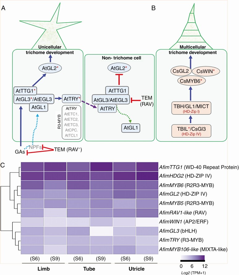
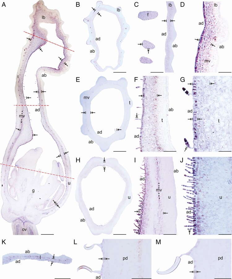
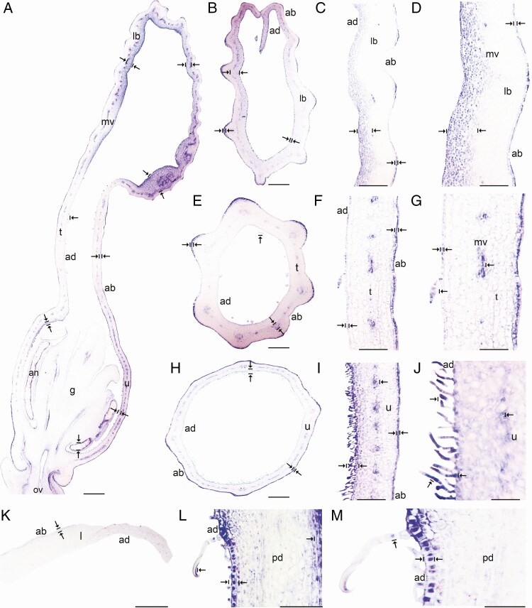
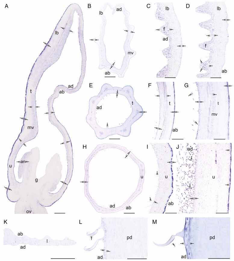
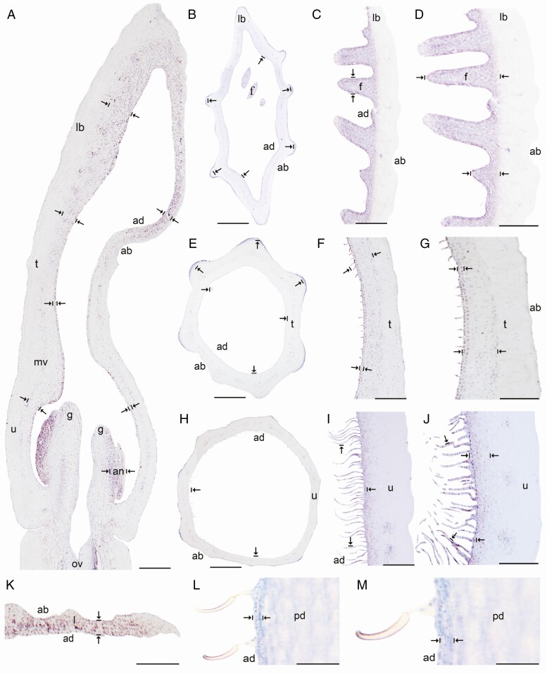
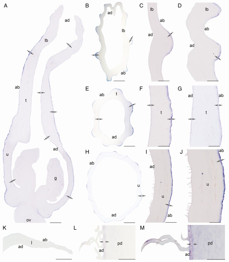
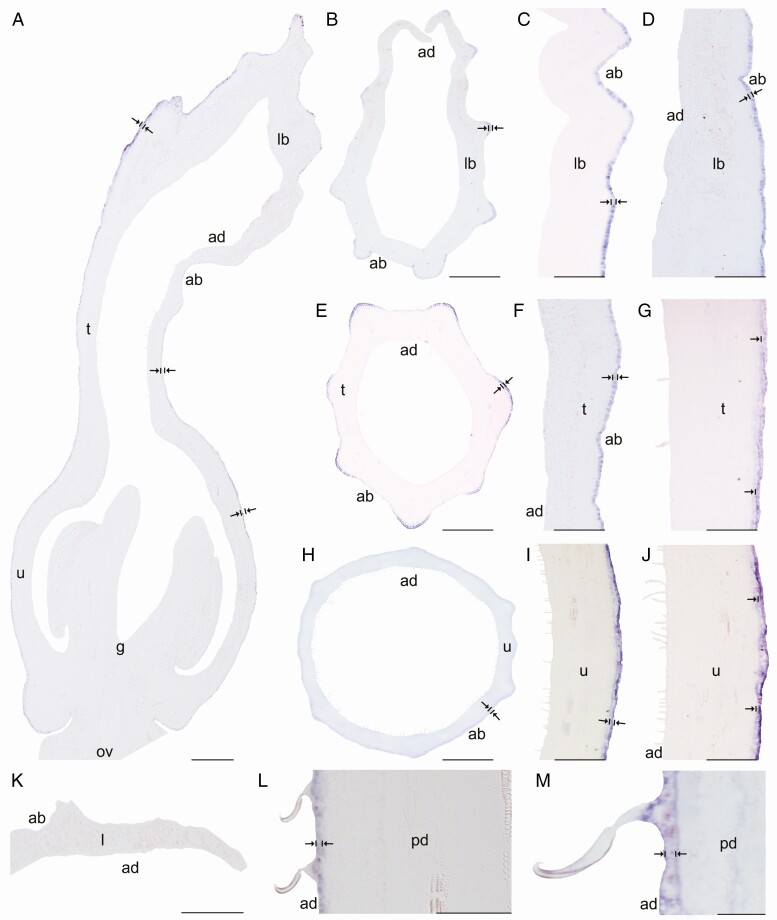
Methods: In this study, we assess the spatio-temporal expression patterns of GL2, GL3, TTG1, TRY, MYB5, MYB6, HDG2, MYB106-like, WIN1 and RAV1-like homologues in the magnoliid Aristolochia fimbriata (Aristolochiaceae) by using comparative RNA-sequencing and in situ hybridization assays.

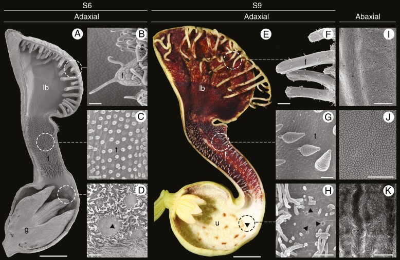
Key results: Genes involved in Aristolochia fimbriata trichome development vary depending on the organ where they are formed. Stem, leaf and pedicel trichomes recruit most of the transcription factors (TFs) described above. Conversely, floral trichomes only use a small subset of genes including AfimGL2, AfimRAV1-like, AfimWIN1, AfimMYB106-like and AfimHDG2. The remaining TFs, AfimTTG1, AfimGL3, AfimTRY, AfimMYB5 and AfimMYB6, are restricted to the abaxial (outer) and the adaxial (inner) pavement epidermal cells.

Conclusions: We re-evaluate the core genetic network shaping trichome fate in flowers of an early-divergent angiosperm lineage and show a morphologically diverse output with a simpler genetic mechanism in place when compared to the models Arabidopsis thaliana and Cucumis sativus. In turn, our results strongly suggest that the canonical trichome gene expression appears to be more conserved in vegetative than in floral tissues across angiosperms.

Keywords: Aristolochia; Piperales; epidermis development; multicellular trichomes; perianth; petaloid sepals.


Figures

Fig. 1.
Fig. 1.
Aristolochia fimbriata, flower morphology and epidermal differentiation of the perianth. (A and E) Sagittal section through flowers at S6 (A) and S9 (E) stages. (B and F) Detail of fimbriae at S6 (B) and S9 (F); note hooked trichomes. (C and G) Secretory multicellular, conical trichomes on the adaxial epidermis of the tube at S6 (C) and S9 (G). (D and H) Detail of the utricle adaxial epidermis with osmophores surrounded by secretory, filamentous multicellular trichomes. (I–K) Detail of the abaxial epidermis of the limb (I), tube (J) and utricle (K), with scattered stomata. Arrowheads: osmophores; f, fimbriae; g, gynostemium; lb, limb; t, tube; u, utricle. Scale bars: 1 mm (A); 100 µm (B–D, I, K); 5 mm (E); 50 µm (F–H, J).
Fig. 2.
Fig. 2.
Canonical signalling pathways for unicellular and multicellular trichome development and comparative expression in Aristolochia fimbriata. (A) Schematic model of Arabidopsis trichome differentiation and surrounding pavement cell fate specification (modified from Zhao et al., 2008; Matías-Hernández et al., 2016). The activation complex is formed by GL1, TTG1 and GL3/EGL3 proteins. This complex activates transcription of GL2 to promote trichome formation while an R3-MYB repressor protein (TRY) moves to the neighbouring cells to repress trichome formation. R3-MYB members including TRY, CPC, ETC1, 2, 3 and TCL1 repress trichome initiation. RAV genes repress trichome initiation via gibberellins. (B) Proposed model for multicellular trichome patterning in Cucumis sativus (modified from Liu et al., 2016). Asterisks indicate the homologues evaluated in this study. (C) Heatmap displaying the expression of the ten candidate genes selected from the two models above. The relative abundance of transcript per million (TPM) values is shown for AfimGL2, AfimGL3, AfimHDG2, AfimMYB5, AfimMYB6, AfimMYB106-like, AfimRAV1-like, AfimTRY, AfimTTG1 and AfimWIN1 in the three perianth portions at S6 and S9.
Fig. 3.
Fig. 3.
In situ hybridization of AfimGL2. (A) Sagittal section of the Aristolochia fimbriata flower at S6; note low expression in the abaxial epidermis with respect to the adaxial epidermis. (B–D) Transverse (B) and longitudinal (C, D) sections of the limb at low (B), mid- (C) and high (D) magnification. (E–G) Transverse (E) and longitudinal (F, G) sections of the tube at low (E), mid- (F) and high (G) magnification. (H–J) Transverse (H) and longitudinal (I, J) sections of the utricle at low (H), mid- (I) and high (J) magnification. (K) Transverse section of the leaf. (L–M) Longitudinal section of the floral pedicel and detail of its hooked trichomes (M) adaxial (ad) of the pedicel here and in Figs 4–8 is given in reference to the shoot axis. Arrow/bar signs point to tissue with positive expression; ab, abaxial epidermis; ad, adaxial epidermis; fi, fimbriae; g, gynostemium; l, leaf; lb, limb; mv, midvein; ov, ovary; pd, pedicel; t, tube; u, utricle. Scale bars: 100 µm (A, B, E, H); 150 µm (C, F, I); 200 µm (D, G, J); 60 µm (K–M).
Fig. 4.
Fig. 4.
In situ hybridization of AfimMYB106-like gene. (A) Sagittal section of the Aristolochia fimbriata flower at S6; note strong expression in the adaxial limb mesophyll and the abaxial epidermis. (B–D) Transverse (B) and longitudinal (C, D) sections of the limb at low (B), mid- (C) and high (D) magnification. (E–G) Transverse (E) and longitudinal (F, G) sections of the tube at low (E), mid- (F) and high (G) magnification. (H–J) Transverse (H) and longitudinal (I–J) sections of the utricle at low (H), mid- (I) and high (J) magnification. (K) Transverse section of the leaf. (L–M) Longitudinal section of the floral pedicel and detail of its hooked trichomes (M). Arrow/bar signs point to tissue with positive expression; ab, abaxial epidermis; ad, adaxial epidermis; an, anther; fi, fimbriae; g, gynostemium; l, leaf; lb, limb; mv, midvein; ov, ovary; pd, pedicel; t, tube; u, utricle. Scale bars: 100 µm (A, B, E, H); 150 µm (C, F, I); 200 µm (D, G, J); 60 µm (K–M).
Fig. 5.
Fig. 5.
In situ hybridization of AfimWIN1. (A) Sagittal section of the Aristolochia fimbriata flower at S6; note higher expression in the adaxial perianth mesophyll, the fimbriae and the abaxial epidermis of the perianth. (B–D) Transverse (B) and longitudinal (C, D) sections of the limb at low (B), mid- (C) and high (D) magnification. (E–G) Transverse (E) and longitudinal (F, G) sections of the tube at low (E), mid- (F) and high (G) magnification. (H–J) Transverse (H) and longitudinal (I–J) sections of the utricle at low (H), mid- (I) and high (J) magnification. (K) Transverse section of the leaf. (L–M) Longitudinal section of the floral pedicel (L) and detail of its hooked trichomes (M). Arrow/bar signs point to tissue with positive expression; ab, abaxial epidermis; ad, adaxial epidermis; an, anther; fi, fimbriae; g, gynostemium; l, leaf; lb, limb; mv, midvein; ov, ovary; pd, pedicel; t, tube; u, utricle. Scale bars: 100 µm (A, B, E, H); 150 µm (C, F, I); 200 µm (D, G, J); 60 µm (K–M).
Fig. 6.
Fig. 6.
In situ hybridization of AfimRAV1-like. (A) Sagittal section of the Aristolochia fimbriata flower at S6; note expression restricted to the adaxial mesophyll of the perianth. (B–D) Transverse (B) and longitudinal (C, D) sections of the limb at low (B), mid- (C) and high (D) magnification. (E–G) Transverse (E) and longitudinal (F, G) sections of the tube at low (E), mid- (F) and high (G) magnification. (H–J) Transverse (H) and longitudinal (I–J) sections of the utricle at low (H), mid- (I) and high (J) magnification. (K) Transverse section of the leaf. (L–M) Longitudinal section of the floral pedicel (L) and detail of its hooked trichomes (M). Arrow/bar signs point to detailed expression patterns; ab, abaxial epidermis; ad, adaxial epidermis; an, anther; fi, fimbriae; g, gynostemium; l, leaf; lb, limb; mv, midvein; ov, ovary; pd, pedicel; t, tube; u, utricle. Scale bars: 100 µm (A, B, E, H); 150 µm (C, F, I); 200 µm (D, G, J); 60 µm (K–M).
Fig. 7.
Fig. 7.
In situ hybridization of AfimHDG2. (A) Sagittal section of the Aristolochia fimbriata flower at S6; note expression in the abaxial epidermis of the perianth, and weak expression in its adaxial counterpart. (B–D) Transverse (B) and longitudinal (C, D) sections of the limb at low (B), mid- (C) and high (D) magnification. (E–G) Transverse (E) and longitudinal (F, G) sections of the tube at low (E), mid- (F) and high (G) magnification. (H–J) Transverse (H) and longitudinal (I–J) sections of the utricle at low (H), mid- (I) and high (J) magnification. (K) Transverse section of the leaf. (L–M) Longitudinal section of the floral pedicel (L) and detail of its the hooked trichomes (M). Arrow/bar signs point to detailed expression patterns; ab, abaxial epidermis; ad, adaxial epidermis; fi, fimbriae; g, gynostemium; l, leaf; lb, limb; mv, midvein; ov, ovary; pd, pedicel; t, tube; u, utricle. Scale bars: 100 µm (A, B, E, H); 150 µm (C, F, I); 200 µm (D, G, J); 60 µm (K–M).
Fig. 8.
Fig. 8.
In situ hybridization of AfimTTG1. (A) Sagittal section of the Aristolochia fimbriata flower at S6; note expression restricted to the abaxial epidermis of the perianth. (B–D) Transverse (B) and longitudinal (C, D) sections of the limb at low (B), mid- (C) and high (D) magnification. (E–G) Transverse (E) and longitudinal (F, G) sections of the tube at low (E), mid- (F) and high (G) magnification. (H–J) Transverse (H) and longitudinal (I, J) sections of the utricle at low (H), mid- (I) and high (J) magnification. (K) Transverse section of the leaf. (L and M) Longitudinal section of the floral pedicel (L) and detail of its hooked trichomes (M). Arrow/bar signs point to detailed expression patterns; ab, abaxial epidermis; ad, adaxial epidermis; fi, fimbriae; g, gynostemium; l, leaf; lb, limb; mv, midvein; ov, ovary; pd, pedicel; t, tube; u, utricle. Scale bars: 100 µm (A, B, E, H); 150 µm (C, F, I); 200 µm (D, G, J); 60 µm (K–M).

Similar articles

Cited by

References

    1. Aliscioni SS, Achler AP, Torretta JP. 2017. Floral anatomy, micromorphology and visitor insects in three species of Aristolochia L. (Aristolochiaceae). New Zealand Journal of Botany 55: 496–513.
    1. Ambrose BA, Lerner DR, Ciceri P, Padilla CM, Yanofsky MF, Schmidt RJ. 2000. Molecular and genetic analyses of the Silky1 gene reveal conservation in floral organ specification between eudicots and monocots. Molecular Cell 5: 569–579. - PubMed
    1. Balkunde R, Pesch M, Hülskamp M. 2011. Trichome patterning in Arabidopsis thaliana: from genetic to molecular models. Current Topics in Developmental Biology 91: 299–321. - PubMed
    1. Baumann K, Perez-Rodriguez M, Bradley D, et al. . 2007. Control of cell and petal morphogenesis by R2R3 MYB transcription factors. Development (Cambridge, England) 134: 1691–1701. - PubMed
    1. Bernhardt C, Lee MM, Gonzalez A, Zhang F, Lloyd A, Schiefelbein J. 2003. The bHLH genes GLABRA3 (GL3) and ENHANCER OF GLABRA3 (EGL3) specify epidermal cell fate in the Arabidopsis root. Development (Cambridge, England) 130: 6431–6439. - PubMed

Publication types

Substances

LinkOut - more resources