DNA methylation-based signatures classify sporadic pituitary tumors according to clinicopathological features
- PMID: 33631002
- PMCID: PMC8328022
- DOI: 10.1093/neuonc/noab044
DNA methylation-based signatures classify sporadic pituitary tumors according to clinicopathological features
Abstract
Background: Distinct genome-wide methylation patterns cluster pituitary neuroendocrine tumors (PitNETs) into molecular groups associated with specific clinicopathological features. Here we aim to identify, characterize, and validate methylation signatures that objectively classify PitNET into clinicopathological groups.
Methods: Combining in-house and publicly available data, we conducted an analysis of the methylome profile of a comprehensive cohort of 177 tumors (Panpit cohort) and 20 nontumor specimens from the pituitary gland. We also retrieved methylome data from an independent PitNET cohort (N = 86) to validate our findings.
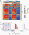
Results: We identified three methylation clusters associated with adenohypophyseal cell lineages and functional status using an unsupervised approach. Differentially methylated probes (DMP) significantly distinguished the Panpit clusters and accurately assigned the samples of the validation cohort to their corresponding lineage and functional subtypes memberships. The DMPs were annotated in regulatory regions enriched with enhancer elements, associated with pathways and genes involved in pituitary cell identity, function, tumorigenesis, and invasiveness. Some DMPs correlated with genes with prognostic and therapeutic values in other intra- or extracranial tumors.
Conclusions: We identified and validated methylation signatures, mainly annotated in enhancer regions that distinguished PitNETs by distinct adenohypophyseal cell lineages and functional status. These signatures provide the groundwork to develop an unbiased approach to classifying PitNETs according to the most recent classification recommended by the 2017 WHO and to explore their biological and clinical relevance in these tumors.
Keywords: DNA methylation; classification; enhancers; pituitary neuroendocrine tumors.
© The Author(s) 2021. Published by Oxford University Press on behalf of the Society for Neuro-Oncology. All rights reserved. For permissions, please e-mail: journals.permissions@oup.com.
Figures


References
-
- Mete O, Cintosun A, Pressman I, Asa SL. Epidemiology and biomarker profile of pituitary adenohypophysial tumors. Mod Pathol. 2018;31(6):900–909. - PubMed
-
- Asa SL, Casar-Borota O, Chanson P, et al. ; attendees of 14th Meeting of the International Pituitary Pathology Club, Annecy, France, November 2016 . From pituitary adenoma to pituitary neuroendocrine tumor (PitNET): an International Pituitary Pathology Club proposal. Endocr Relat Cancer. 2017;24(4):C5–C8. - PubMed
-
- Molitch ME. Diagnosis and treatment of pituitary adenomas: a review. JAMA. 2017;317(5):516–524. - PubMed
-
- Mete O, Lopes MB. Overview of the 2017 WHO classification of pituitary tumors. Endocr Pathol. 2017;28(3):228–243. - PubMed
Publication types
MeSH terms
Grants and funding
LinkOut - more resources
Full Text Sources
Other Literature Sources
Medical

