

Comment
doi: 10.1016/j.bbi.2021.02.013.
Epub 2021 Feb 23.
Indirect immunofluorescence for detecting anti-neuronal autoimmunity in CSF after COVID-19 - Possibilities and pitfalls
Affiliations
- PMID: 33631284
- PMCID: PMC9761871
- DOI: 10.1016/j.bbi.2021.02.013
Item in Clipboard
Comment
Indirect immunofluorescence for detecting anti-neuronal autoimmunity in CSF after COVID-19 - Possibilities and pitfalls
Brain Behav Immun.
2021 May.
No abstract available
Figures
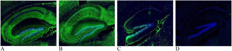
Images are of 16 μm paraformaldehyde (4%) fixed sagittal cryosections of mouse hippocampus. Immunohistochemistry was performed using cerebrospinal fluid (CSF) diluted 1:4 to detect anti-neuronal antibodies (green; cell nuclei marker DAPI is blue). A: CSF from a patient with COVID-19 associated acute necrotizing encephalopathy where neuropil staining is found in the subiculum, stratum oriens and stratum radiatum in all cornu ammonis (CA) subfields, dentate gyrus polymorph layer and faint in the molecular layer. The pyramidal layer of CA1 and stratum lancosum-moleculare are both negative. B: CSF from a patient presenting with COVID-19 associated acute hemorrhagic leukoencephalitis. In the hippocampus and the cortex (see top left corner) there is a general dendritic staining. Staining can also be observed in some specific cell somas, especially in the subiculum and cortex (not shown). The signal is fainter in the CA1 stratum oriens, CA1 stratum lancosum and the dentate gyrus polymorph layer compared to the rest of the area. C: CSF from patient with COVID-19 associated malignant catatonia shows membrane staining of cells in the pyramidal layer, subiculum and granule cell layer of the dentate gyrus. D: Reference CSF sample, from a donor with bipolar disease without suspected autoimmunity.
Comment in
-
Letter to the Editor: Comment on Mulder J et al. (2021) indirect immunofluorescence for detecting anti-neuronal autoimmunity in CSF after COVID-19 - possibilities and pitfalls.Brain Behav Immun. 2021 May;94:475. doi: 10.1016/j.bbi.2021.02.014. Epub 2021 Feb 25. Brain Behav Immun. 2021. PMID: 33639240 Free PMC article. No abstract available.
Comment on
-
High frequency of cerebrospinal fluid autoantibodies in COVID-19 patients with neurological symptoms.Brain Behav Immun. 2021 Mar;93:415-419. doi: 10.1016/j.bbi.2020.12.022. Epub 2020 Dec 24. Brain Behav Immun. 2021. PMID: 33359380 Free PMC article.
References
-
- Franke C., Ferse C., Kreye J., Reincke S.M., Sanchez-Sendin E., Rocco A., Steinbrenner M., Angermair S., Treskatsch S., Zickler D., Eckardt K.-U., Dersch R., Hosp J., Audebert H.J., Endres M., Ploner J.C., Prüß H. High frequency of cerebrospinal fluid autoantibodies in COVID-19 patients with neurological symptoms. Brain Behav. Immun. 2020 doi: 10.1016/j.bbi.2020.12.022. - DOI - PMC - PubMed
-
- Virhammar J., Kumlien E., Fällmar D., Frithiof R., Jackmann S., Sköld M.K., Kadir M., Frick J., Lindeberg J., Olivero-Reinius H., Ryttlefors M., Cunningham J.L., Wikström J., Grabowska A., Bondeson K., Bergquist J., Zetterberg H., Rostami E. Acute necrotizing encephalopathy with SARS-CoV-2 RNA confirmed in cerebrospinal fluid. Neurology. 2020;95(10):445–449. doi: 10.1212/WNL.0000000000010250. - DOI - PMC - PubMed
-
- Mulder J., Feresiadou A., Fallmar D., Frithiof R., Virhammar J., Rasmusson J.A., Rostami E., Kumlien E., Cunningham L.J. Autoimmune encephalitis presenting with acute excited catatonia in a 40-year-old male patient with Covid-19. Am. J. Psychiatry. 2020 doi: 10.1176/appi.ajp.2020.20081236. In press. - DOI - PubMed
-
- Sjostedt, E., Zhong, W., Fagerberg, L., et al. An atlas of the protein-coding genes in the human, pig, and mouse brain. Science. 2020;367(6482). doi: 10.1126/science.aay5947. - PubMed
Publication types
MeSH terms
Substances
LinkOut - more resources
Full Text Sources
Other Literature Sources
Medical
