Review
. 2022;20(1):223-253.
doi: 10.2174/1570159X19666210225152728.

Central Neural Circuits Orchestrating Thermogenesis, Sleep-Wakefulness States and General Anesthesia States

Affiliations
Review

Central Neural Circuits Orchestrating Thermogenesis, Sleep-Wakefulness States and General Anesthesia States

Jiayi Wu et al. Curr Neuropharmacol. 2022.

Abstract

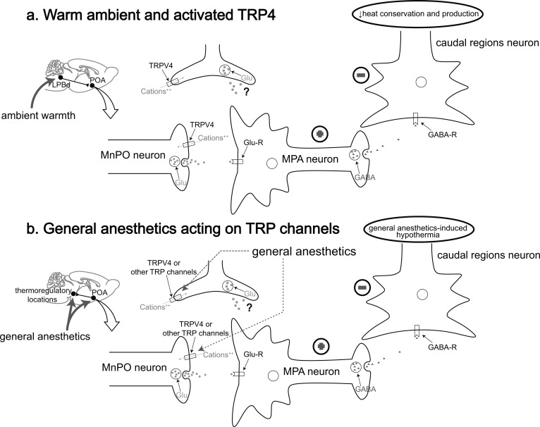
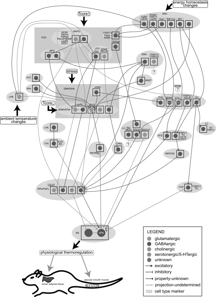
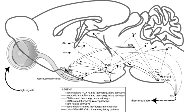
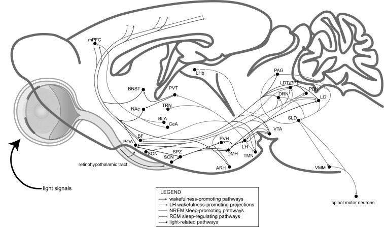
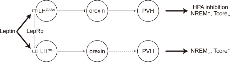
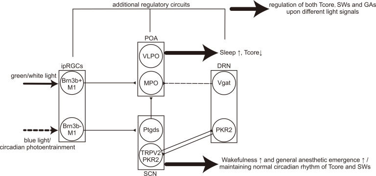
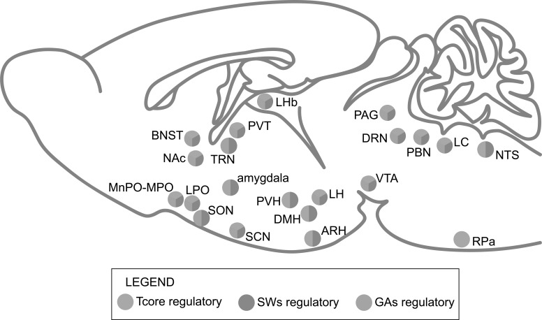
Great progress has been made in specifically identifying the central neural circuits (CNCs) of the core body temperature (Tcore), sleep-wakefulness states (SWs), and general anesthesia states (GAs), mainly utilizing optogenetic or chemogenetic manipulations. We summarize the neuronal populations and neural pathways of these three CNCs, which gives evidence for the orchestration within these three CNCs, and the integrative regulation of these three CNCs by different environmental light signals. We also outline some transient receptor potential (TRP) channels that function in the CNCs-Tcore and are modulated by some general anesthetics, which makes TRP channels possible targets for addressing the general-anestheticsinduced- hypothermia (GAIH). We suggest this review will provide new orientations for further consummating these CNCs and elucidating the central mechanisms of GAIH.

Keywords: Central neural circuits; TRP channels; body temperature regulation; chemogenetics; general anesthesia; intrinsically photosensitive retinal ganglion cells; optogenetics; sleep-wakefulness states.


Figures

Fig. (1)
Fig. (1)
A model of TRPV4 and other possible TRP channels functioning in the central thermoregulatory pathway under warm ambient and general anesthesia. (a) Ambient warmth activates the LPBd→MnPO pathway, subsequently activating TRPV4 located in the synaptic terminals of the MnPO and causing an influx of cations to contribute to the glutamate release from these neurons. Then the postsynaptic GABAergic MPA neurons are activated (indicated by “plus sign” signal), thus inhibiting the neurons in caudal regions (indicated by ”minus sign” signal) to cause a reduction in heat conservation and production. Alternatively, TRPV4 may present in the astrocytes and regulate these thermoregulatory responses. (b) Under sedation or general anesthesia, the POA as well as other possible central thermoregulatory locations is activated by general anesthetics, TRPV4 or other possible TRP channels, such as heat-activated TRPV1 and cold-activated TRPM8, may be the target for general anesthetics. Similar as the procedures represented above, general anesthetics may activate the heat-activated TRP channels or inhibit the cold-activated TRP channels (not shown in this model), thereby causing hypothermia. LPBd, the dorsal part of lateral parabrachial nucleus; Glu, glutamate; Glu-R, glutamate receptor; GABA-R, GABA receptor. (Adapted from [74]).
Fig. (2)
Fig. (2)
A schematic of the specific central thermoregulatory neuronal populations and neural pathways. The signals of ambient temperature changes, energy homeostasis changes and stress are input from the LPB, ARH and DMH/DHA, respectively. The POA is heat-activated and the dDMH/DHA is cold-activated. The glutamatergic MnPO neurons reported to co-express some specific cell type markers (presented in the yellow box) send projections to some downstream locations, including the LPO, dDMH/DHA, rRPa/PaPy and PVH, while the GABAergic MnPO neurons locally inhibit the MPO and LPO. The Brs3+ dDMH/DHA neurons, which receive inputs from the POA (uncharted), vDMH, ARH, BNST, PVT, NAc and RIP/RMg, are glutamatergic and project to the rRPa/PaPy, together with the dDMH/DHALepRb→rRPa/PaPy→sympathetic preganglionic neurons (SPN), dDMH/DHAAch→rRPa/PaPy5-HT→spinal intermediolateral nucleus (IML) and dDMH/DHA→PAG pathways. The rRPa/PaPy may also receive projections from LPOVglut2 neurons (uncharted) and the LHbGABA innervating VTA neurons which are proposed to be dopaminergic or serotonergic. The metabolism-related ARH send inhibitory projections to the LPB, POA and PVH, thus modulating the canonical thermoregulatory circuits and regulating the neuroendocrine outputs from Crh+ PVHmpv neurons to the orexin neurons or NTSGABA neurons. PVHTH neurons project to downstream NTS, LC and VLM. PVHmpdBDNF neurons, receiving projections from ARHAgRP and ARHPOMC neurons, directly project to the SPN. The PVH is also regulated by the stress signals input from the vDMH. The IRt/PCRtGABA neurons in the downstream hypothalamic neuropeptide Y (NPY)-related pathways inhibit the rRPa/PaPy and subsequently the BAT thermogenesis. In the LH, receiving projections from TRPM8+ POA neurons, the orexin neurons project to the rRPa/PaPy and the Bdnf-e1 glutamatergic neurons directly innervate the SPN. The DRN is another important thermoregulatory location with GABAergic projections to the MPO, dDMH/DHA, rRPa/PaPy and BNST. The DRN5-HT neurons proposed to co-express TRPV3/TRPV4 may also be involved in thermoregulation. All these pathways cooperatively regulate the physiological thermo-responses which especially characterized by the brown adipose tissue (BAT) thermogenesis and tail vasomotor responses. Tcore, core body temperature; the question mark in the yellow box means that cell type marker is uncertain.
Fig. (3)
Fig. (3)
A model of the central thermoregulatory circuits controlled by environmental light signals. The light signal as well as the circadian photoentrainment is transmitted to the SCN and POA through the retinohypothalamic tract and then the downstream subparaventricular zone (SPZ), DMH and PVH. The POA→DMH→RPa/VLM pathway is canonical, functioning together with other thermoregulatory circuits, including the ARH-, DMH-, DRN-, osmo-sodium-related and other possible pathways.
Fig. (4)
Fig. (4)
A model of the central neural circuits of sleep-wakefulness states controlled by environmental light signals. The light-related pathway also regulates the LH and LC, both of which are wakefulness-promoting. The LH sends innervations to extensive locations, including the basal forebrain (BF), TRN, PVT, TMN, VTA, DRN, laterodorsal tegmental area (LDT)/ pedunculopontine tegmental area (PPT), PBN and LC. The arousal-promoting system, includes the LC, PBN, LDT/PPT, DRN, VTA, TMN, BF, PVT, NAc and BNST, while the NREM sleep-promoting pathways involves the inhibition of the wakefulness-promoting pathways by the POA, the inhibition of the cortex by the basal forebrain (BF), and the inhibition of the LH by VTA. The LHb is also NREM sleep-promoting, while the related pathway(s) is not clear (indicated by dotted line). The DRN can be wakefulness-promoting when hungry while sleep-permissive during satiety. Additionally, the latest finding suggests that the VLPOVglut2 neurons are wakefulness-promoting whilst the MnPOVgat neurons are NREM sleep-promoting. The DRN, PAG and LC participate in the REM sleep-regulating pathways to mutually regulate the muscle paralysis of REM sleep by directly inhibiting the excitatory sublaterodorsal nucleus (SLD)→VMM pathway or indirectly inhibit the excitatory LDT/PPT→SLD pathway, and the PAG receives inhibitory projections from the SLD. The LDT/PPT→BF pathway may help drive the typical fast EEG activity of REM sleep. And the BLA/CeA→medial prefrontal cortex (mPFC) pathway may also regulate REM sleep. (Adapted from [8]).
Fig. (5)
Fig. (5)
A possible model of leptin signals co-regulating core body temperature and sleep-wakefulness states. The leptin signals activate the LepRb of both the LHGABA neurons and LHNts neurons, with the former inhibit the orexin neurons that innervate the PVH to subsequently induce inhibition of the hypothalamic-pituitary-adrenal (HPA), increase of NREM sleep and decrease of Tcore, while the latter contrarily activate the orexin neurons and downstream location(s), possibly the PVH (indicated by dotted arrow), to reduce NREM sleep and elevate Tcore.
Fig. (6)
Fig. (6)
A model of central neural circuits regulated by the acute light exposure/circadian photoentrainment. The acute green/white light stimuli may be transmitted by the Brn3b+ ipRGCs→POA pathway to promote sleep and decrease Tcore, while the blue light may stimulate the Brn3b- ipRGCs→SCN pathway to promote wakefulness as well as emergence from general anesthesia. The circadian rhythm of Tcore and SWs may also be regulated by the Brn3b- ipRGCs→SCN pathway. The SCN neurons that co-express TRPV2 and PKR2 are mutually innervated with the PKR2+ DRN neurons to regulate the circadian rhythm. The Ptgds+ SCN neurons may release thermoregulatory PGD2 to the POA to regulate the circadian oscillation of Tcore. The DRNVgat neurons inhibit the MPO neurons to modulate Tcore. Together with other regulatory circuits, different light signals are able to regulate Tcore, SWs and general anesthetic states (GAs). Full lines represent light-related/circadian projections and dotted line represents simplex thermoregulatory projection.
Fig. (7)
Fig. (7)
Evidence for central orchestration among core body temperature, sleep-wakefulness states and general anesthesia states. A sagittal view of the rodent brain shows the approximate locations of the nuclei known to play a role in regulating Tcore and(or) SWs, which may also be associated with regulation of GAs.

Similar articles

References

    1. Latifi B., Adamantidis A., Bassetti C., Schmidt M.H. Sleep-wake cycling and energy conservation: role of hypocretin and the lateral hypothalamus in dynamic state-dependent resource optimization. Front. Neurol. 2018;9:790. doi: 10.3389/fneur.2018.00790. - DOI - PMC - PubMed
    1. Harding E.C., Franks N.P., Wisden W. The temperature dependence of sleep. Front. Neurosci. 2019;13:336. doi: 10.3389/fnins.2019.00336. - DOI - PMC - PubMed
    1. Morrison S.F., Nakamura K. Central mechanisms for thermoregulation. Annu. Rev. Physiol. 2019;81:285–308. doi: 10.1146/annurev-physiol-020518-114546. - DOI - PubMed
    1. Tan C.L., Knight Z.A. Regulation of body temperature by the nervous system. Neuron. 2018;98(1):31–48. doi: 10.1016/j.neuron.2018.02.022. - DOI - PMC - PubMed
    1. Morrison S.F., Madden C.J., Tupone D. Central neural regulation of brown adipose tissue thermogenesis and energy expenditure. Cell Metab. 2014;19(5):741–756. doi: 10.1016/j.cmet.2014.02.007. - DOI - PMC - PubMed