

Treatment with endoscopic transnasal resection of hypothalamic pilocytic astrocytomas: a single-center experience
- PMID: 33632188
- PMCID: PMC7908641
- DOI: 10.1186/s12893-021-01113-6
Treatment with endoscopic transnasal resection of hypothalamic pilocytic astrocytomas: a single-center experience
Abstract
Backgrounds: Pilocytic astrocytomas (PAs) are World Health Organization (WHO) grade I tumors, which are relatively common, and are benign lesions in children. PAs could originate from the cerebellum, optic pathways, and third ventricular/hypothalamic region. Traditional various transcranial routes are used for hypothalamic PAs (HPAs). However, there are few studies on hypothalamic PAs treated through the endoscopic endonasal approach (EEA). This study reports the preliminary experience of the investigators and results with HPAs via expanded EEAs.
Methods: All patients with HPAs, undergone EEA in our hospital from 2017 to 2019, were retrospectively reviewed. The demographic data, clinical symptoms, complications, skull base reconstruction, prognosis, and endocrinological data were all recorded and analyzed in detail.
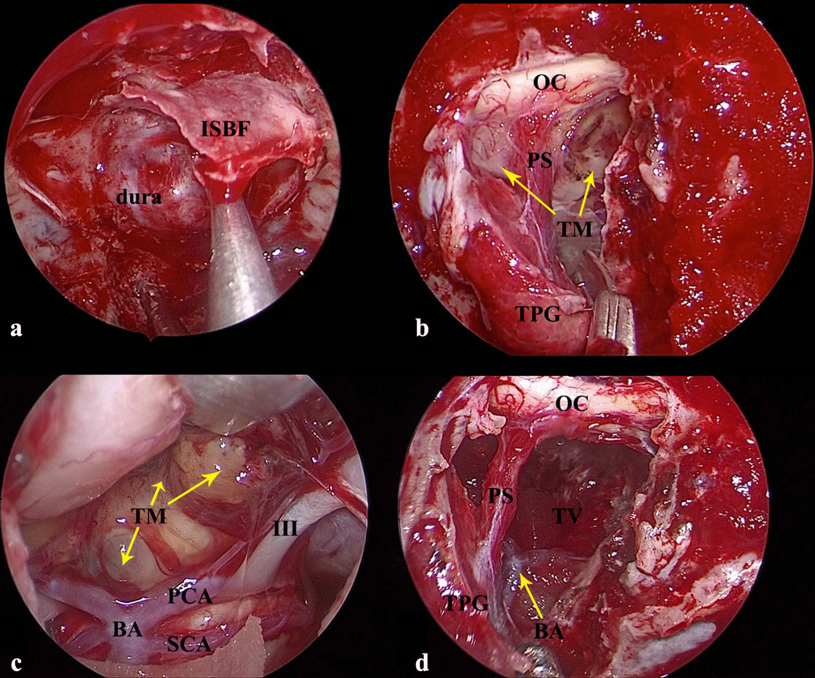
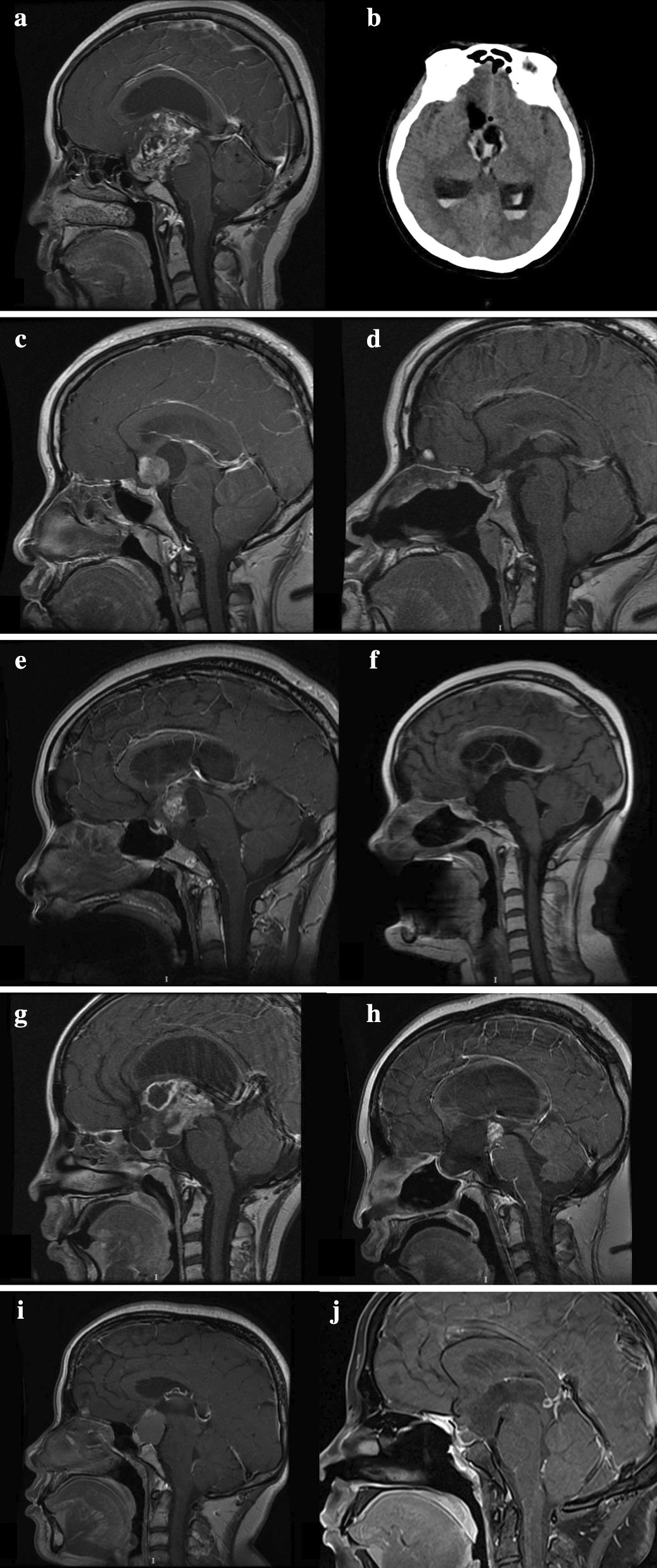
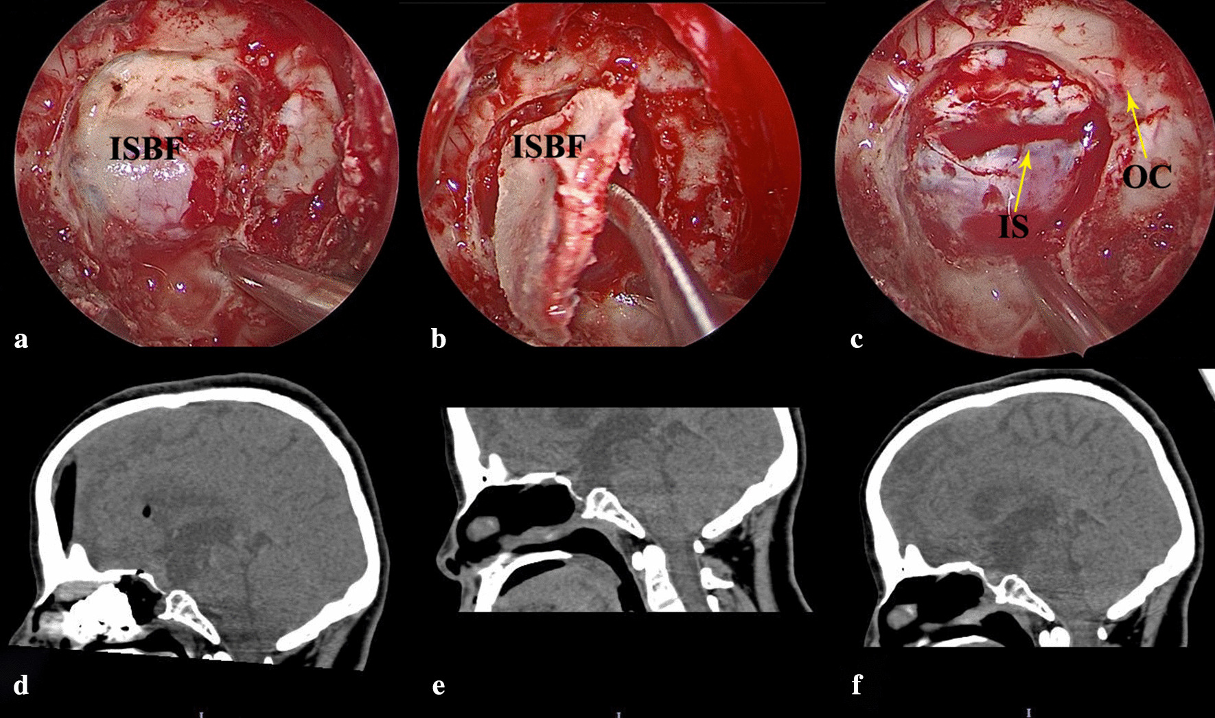
Results: Finally, five female patients were enrolled. The average age of patients was 28.6 ± 14.0. All patients had complaints about their menstrual disorder. One patient had severe bilateral visual impairment. Furthermore, only one patient suffered from severe headache due to acute hydrocephalus, although there were four patients with headache or dizziness. Four cases achieved gross-total resection, and one patient achieved subtotal resection. Furthermore, there was visual improvement in one patient (case 5), and postoperative worsening of vision in one patient (case 4). However, only one patient had postoperative intracranial infection. None of the patients experienced a postoperative CSF leak, and in situ bone flap (ISBF) techniques were used for two cases for skull base repair. In particular, ISBF combined with free middle turbinate mucosal flap was used for case 5. After three years of follow-up, three patients are still alive, two patients had no neurological or visual symptoms, or tumor recurrence, and one patient had severe hypothalamic dysfunction. Unfortunately, one patient died of severe postoperative hypothalamus reaction, which presented with coma, high fever, diabetes insipidus, hypernatremia and intracranial infection. The other patient died of recurrent severe pancreatitis at one year after the operation.
Conclusion: Although the data is still very limited and preliminary, EEA provides a direct approach to HPAs with acceptable prognosis in terms of tumor resection, endocrinological and visual outcomes. ISBF technique is safe and reliable for skull base reconstruction.
Keywords: Endoscopic transnasal resection; Hypothalamic; In situ bone flap; Pilocytic astrocytomas.
Conflict of interest statement
The authors declare that they have no competing interests.
Figures
Similar articles
-
Endoscopic transnasal resection of optic pathway pilocytic astrocytoma.Childs Nerv Syst. 2019 Jan;35(1):73-81. doi: 10.1007/s00381-018-3994-4. Epub 2018 Oct 18. Childs Nerv Syst. 2019. PMID: 30338361
-
Expanding indications for the extended endoscopic endonasal approach to hypothalamic gliomas: preliminary report.Neurosurg Focus. 2014;37(4):E11. doi: 10.3171/2014.7.FOCUS14317. Neurosurg Focus. 2014. PMID: 25270130
-
Suprasellar and recurrent pediatric craniopharyngiomas: expanding indications for the extended endoscopic transsphenoidal approach.J Neurosurg Pediatr. 2018 Jan;21(1):72-80. doi: 10.3171/2017.7.PEDS17295. Epub 2017 Nov 10. J Neurosurg Pediatr. 2018. PMID: 29125446
-
Endocrinological and ophthalmological consequences of an initial endonasal endoscopic approach for resection of craniopharyngiomas.Neurosurg Focus. 2010 Apr;28(4):E8. doi: 10.3171/2010.1.FOCUS09292. Neurosurg Focus. 2010. PMID: 20367365 Review.
-
Endoscopic transnasal surgery in optic pathway gliomas located in the chiasma-hypothalamic region: case series of ten patients in a single-center experience and endoscopic literature review.Childs Nerv Syst. 2022 Nov;38(11):2071-2082. doi: 10.1007/s00381-022-05665-7. Epub 2022 Sep 10. Childs Nerv Syst. 2022. PMID: 36087131 Review.
Cited by
-
Skull base reconstruction using in situ bone flap in patients with pituitary adenomas treated by endoscopic endonasal approach.Front Neurol. 2023 Jun 14;14:1194251. doi: 10.3389/fneur.2023.1194251. eCollection 2023. Front Neurol. 2023. PMID: 37388547 Free PMC article.
-
Management of Optic Pathway Glioma: A Systematic Review and Meta-Analysis.Cancers (Basel). 2022 Sep 30;14(19):4781. doi: 10.3390/cancers14194781. Cancers (Basel). 2022. PMID: 36230704 Free PMC article. Review.
-
Hypothalamic Hemangioma-like Pilocytic Astrocytoma in an Adult Patient: A Systematic Review with a Focus on Differential Diagnosis and Neurological Presentation.J Clin Med. 2024 Jun 17;13(12):3536. doi: 10.3390/jcm13123536. J Clin Med. 2024. PMID: 38930064 Free PMC article. Review.
References
MeSH terms
LinkOut - more resources
Full Text Sources
Other Literature Sources
