

Niclosamide suppresses the expansion of follicular helper T cells and alleviates disease severity in two murine models of lupus via STAT3
- PMID: 33632240
- PMCID: PMC7908700
- DOI: 10.1186/s12967-021-02760-2
Niclosamide suppresses the expansion of follicular helper T cells and alleviates disease severity in two murine models of lupus via STAT3
Abstract
Background: Autoantibody production against endogenous cellular components is pathogenic feature of systemic lupus erythematosus (SLE). Follicular helper T (TFH) cells aid in B cell differentiation into autoantibody-producing plasma cells (PCs). The IL-6 and IL-21 cytokine-mediated STAT3 signaling are crucial for the differentiation to TFH cells. Niclosamide is an anti-helminthic drug used to treat parasitic infections but also exhibits a therapeutic effect on autoimmune diseases due to its potential immune regulatory effects. In this study, we examined whether niclosamide treatment could relieve lupus-like autoimmunity by modulating the differentiation of TFH cells in two murine models of lupus.
Methods: 10-week-old MRL/lpr mice were orally administered with 100 mg/kg of niclosamide or with 0.5% methylcellulose (MC, vehicle) daily for 7 weeks. TLR7 agonist, resiquimod was topically applied to an ear of 8-week-old C57BL/6 mice 3 times a week for 5 weeks. And they were orally administered with 100 mg/kg of niclosamide or with 0.5% MC daily for 5 weeks. Every mouse was analyzed for lupus nephritis, proteinuria, autoantibodies, immune complex, immune cell subsets at the time of the euthanization.
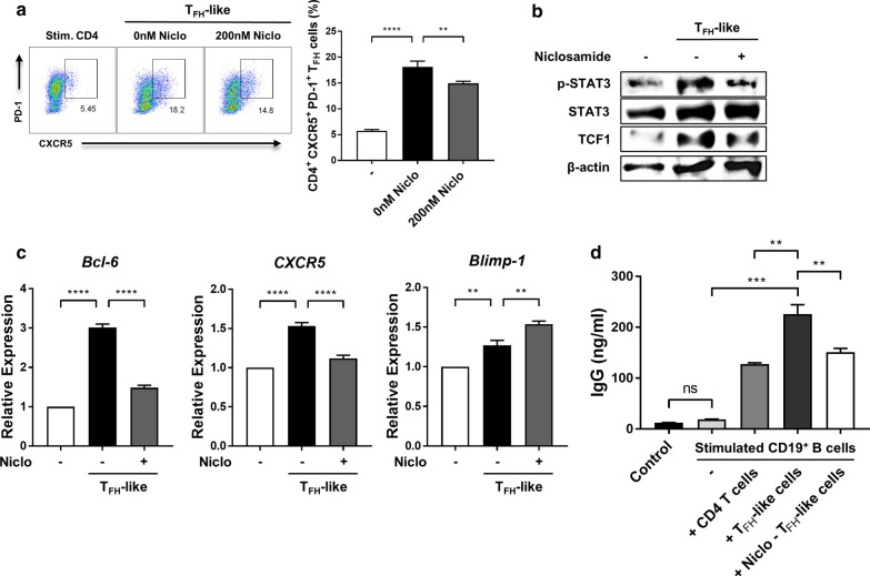
Results: Niclosamide treatment greatly improved proteinuria, anti-dsDNA antibody levels, immunoglobulin subclass titers, histology of lupus nephritis, and C3 deposition in MRL/lpr and R848-induced mice. In addition, niclosamide inhibited the proportion of TFH cells and PCs in the spleens of these animals, and effectively suppressed differentiation of TFH-like cells and expression of associated genes in vitro.
Conclusions: Niclosamide exerted therapeutic effects on murine lupus models by suppressing TFH cells and plasma cells through STAT3 inhibition.
Keywords: Follicular helper T cells; MRL/lpr; Niclosamide; R848-induced model; STAT3; Systemic lupus erythematosus.
Conflict of interest statement
The authors declare that they have no competing interest.
Figures


Similar articles
-
Therapeutic effects of artesunate on lupus-prone MRL/lpr mice are dependent on T follicular helper cell differentiation and activation of JAK2-STAT3 signaling pathway.Phytomedicine. 2019 Sep;62:152965. doi: 10.1016/j.phymed.2019.152965. Epub 2019 May 19. Phytomedicine. 2019. PMID: 31129432
-
Toll-like receptor 7 agonist imiquimod prevents the progression of SLE in MRL/lpr mice via inhibiting the differentiation of T follicular helper cells.Int Immunopharmacol. 2020 Mar;80:106239. doi: 10.1016/j.intimp.2020.106239. Epub 2020 Jan 30. Int Immunopharmacol. 2020. PMID: 32007709
-
Sirtuin 1 overexpression contributes to the expansion of follicular helper T cells in systemic lupus erythematosus and may serve as an accessible therapeutic target.Rheumatology (Oxford). 2024 May 3;63(6):1699-1709. doi: 10.1093/rheumatology/kead453. Rheumatology (Oxford). 2024. PMID: 37665721
-
New insights into the immunopathogenesis of systemic lupus erythematosus: the role of T follicular helper cells.Chin Med J (Engl). 2014;127(19):3496-502. Chin Med J (Engl). 2014. PMID: 25269920 Review.
-
T follicular helper (Tfh) cells in lupus: Activation and involvement in SLE pathogenesis.Eur J Immunol. 2016 Feb;46(2):281-90. doi: 10.1002/eji.201545760. Eur J Immunol. 2016. PMID: 26614103 Review.
Cited by
-
Repurposing Niclosamide as a plausible neurotherapeutic in autism spectrum disorders, targeting mitochondrial dysfunction: a strong hypothesis.Metab Brain Dis. 2024 Mar;39(3):387-401. doi: 10.1007/s11011-023-01247-x. Epub 2023 Jun 7. Metab Brain Dis. 2024. PMID: 37284987 Free PMC article. Review.
-
Niclosamide Alleviated Skin Inflammation and Restored the Balance between Effector and Regulatory T Cells in Skin.Biomol Ther (Seoul). 2025 Jul 1;33(4):735-745. doi: 10.4062/biomolther.2024.210. Epub 2025 Jun 10. Biomol Ther (Seoul). 2025. PMID: 40492423 Free PMC article.
-
Dexamethasone reduces autoantibody levels in MRL/lpr mice by inhibiting Tfh cell responses.J Cell Mol Med. 2021 Sep;25(17):8329-8337. doi: 10.1111/jcmm.16785. Epub 2021 Jul 28. J Cell Mol Med. 2021. PMID: 34318604 Free PMC article.
-
Niclosamide-A promising treatment for COVID-19.Br J Pharmacol. 2022 Jul;179(13):3250-3267. doi: 10.1111/bph.15843. Epub 2022 Apr 11. Br J Pharmacol. 2022. PMID: 35348204 Free PMC article. Review.
-
The differentiation courses of the Tfh cells: a new perspective on autoimmune disease pathogenesis and treatment.Biosci Rep. 2024 Jan 31;44(1):BSR20231723. doi: 10.1042/BSR20231723. Biosci Rep. 2024. PMID: 38051200 Free PMC article. Review.
References
Publication types
MeSH terms
Substances
LinkOut - more resources
Full Text Sources
Other Literature Sources
Medical
Miscellaneous
