

Inflammatory, synaptic, motor, and behavioral alterations induced by gestational sepsis on the offspring at different stages of life
- PMID: 33632243
- PMCID: PMC7905683
- DOI: 10.1186/s12974-021-02106-1
Inflammatory, synaptic, motor, and behavioral alterations induced by gestational sepsis on the offspring at different stages of life
Abstract
Background: The term sepsis is used to designate a systemic condition of infection and inflammation associated with hemodynamic changes that result in organic dysfunction. Gestational sepsis can impair the development of the central nervous system and may promote permanent behavior alterations in the offspring. The aim of our work was to evaluate the effects of maternal sepsis on inflammatory cytokine levels and synaptic proteins in the hippocampus, neocortex, frontal cortex, and cerebellum of neonatal, young, and adult mice. Additionally, we analyzed the motor development, behavioral features, and cognitive impairments in neonatal, young and adult offspring.
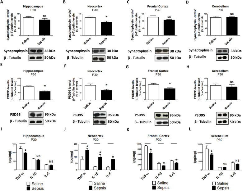
Methods: Pregnant mice at the 14th embryonic day (E14) were intratracheally instilled with saline 0.9% solution (control group) or Klebsiella spp. (3 × 10⁸ CFU) (sepsis group) and started on meropenem after 5 h. The offspring was sacrificed at postnatal day (P) 2, P8, P30, and P60 and samples of liver, lung, and brain were collected for TNF-α, IL-1β, and IL-6 measurements by ELISA. Synaptophysin, PSD95, and β-tubulin levels were analyzed by Western blot. Motor tests were performed at all analyzed ages and behavioral assessments were performed in offspring at P30 and P60.
Results: Gestational sepsis induces a systemic pro-inflammatory response in neonates at P2 and P8 characterized by an increase in cytokine levels. Maternal sepsis induced systemic downregulation of pro-inflammatory cytokines, while in the hippocampus, neocortex, frontal cortex, and cerebellum an inflammatory response was detected. These changes in the brain immunity were accompanied by a reduction of synaptophysin and PSD95 levels in the hippocampus, neocortex, frontal cortex, and cerebellum, in all ages. Behavioral tests demonstrated motor impairment in neonates, and depressive-like behavior, fear-conditioned memory, and learning impairments in animals at P30 and P60, while spatial memory abilities were affected only at P60, indicating that gestational sepsis not only induces an inflammatory response in neonatal mouse brains, but also affects neurodevelopment, and leads to a plethora of behavioral alterations and cognitive impairments in the offspring.
Conclusion: These data suggest that maternal sepsis may be causatively related to the development of depression, learning, and memory impairments in the litter.
Keywords: Depression; Gestational sepsis; Memory; Motor damage; Synaptophysin.
Conflict of interest statement
The authors declare no relevant conflicts of interest.
Figures


Similar articles
-
Gestational hypothyroxinemia induces ASD-like phenotypes in behavior, proinflammatory markers, and glutamatergic protein expression in mouse offspring of both sexes.Front Endocrinol (Lausanne). 2024 May 1;15:1381180. doi: 10.3389/fendo.2024.1381180. eCollection 2024. Front Endocrinol (Lausanne). 2024. PMID: 38752179 Free PMC article.
-
TLR4 response mediates ethanol-induced neurodevelopment alterations in a model of fetal alcohol spectrum disorders.J Neuroinflammation. 2017 Jul 24;14(1):145. doi: 10.1186/s12974-017-0918-2. J Neuroinflammation. 2017. PMID: 28738878 Free PMC article.
-
Maternal immune activation by poly I:C induces expression of cytokines IL-1β and IL-13, chemokine MCP-1 and colony stimulating factor VEGF in fetal mouse brain.J Neuroinflammation. 2012 Apr 30;9:83. doi: 10.1186/1742-2094-9-83. J Neuroinflammation. 2012. PMID: 22546005 Free PMC article.
-
Offspring neuroimmune consequences of maternal malnutrition: Potential mechanism for behavioral impairments that underlie metabolic and neurodevelopmental disorders.Front Neuroendocrinol. 2017 Oct;47:109-122. doi: 10.1016/j.yfrne.2017.07.007. Epub 2017 Jul 20. Front Neuroendocrinol. 2017. PMID: 28736323 Free PMC article. Review.
-
Long-Term Cognitive Outcomes After Sepsis: a Translational Systematic Review.Mol Neurobiol. 2019 Jan;56(1):186-251. doi: 10.1007/s12035-018-1048-2. Epub 2018 Apr 23. Mol Neurobiol. 2019. PMID: 29687346
Cited by
-
Microglia-triggered hypoexcitability plasticity of pyramidal neurons in the rat medial prefrontal cortex.Curr Res Neurobiol. 2022 Feb 5;3:100028. doi: 10.1016/j.crneur.2022.100028. eCollection 2022. Curr Res Neurobiol. 2022. PMID: 36518339 Free PMC article.
-
The biological alterations of synapse/synapse formation in sepsis-associated encephalopathy.Front Synaptic Neurosci. 2022 Dec 2;14:1054605. doi: 10.3389/fnsyn.2022.1054605. eCollection 2022. Front Synaptic Neurosci. 2022. PMID: 36530954 Free PMC article. Review.
-
The Biological Changes of Synaptic Plasticity in the Pathological Process of Sepsis-associated Encephalopathy.Curr Neuropharmacol. 2025;23(4):359-374. doi: 10.2174/1570159X23666241028105746. Curr Neuropharmacol. 2025. PMID: 39473252 Free PMC article. Review.
-
Microglial Priming in Infections and Its Risk to Neurodegenerative Diseases.Front Cell Neurosci. 2022 Jun 15;16:878987. doi: 10.3389/fncel.2022.878987. eCollection 2022. Front Cell Neurosci. 2022. PMID: 35783096 Free PMC article. Review.
-
Utility of melatonin on brain injury, synaptic transmission, and energy metabolism in rats with sepsis.Naunyn Schmiedebergs Arch Pharmacol. 2025 Feb;398(2):1509-1519. doi: 10.1007/s00210-024-03337-8. Epub 2024 Aug 6. Naunyn Schmiedebergs Arch Pharmacol. 2025. PMID: 39105798 Free PMC article.
References
-
- Abraham E. New definitions for sepsis and septic shock continuing evolution but with much still to be done. J Am Med Assoc. 2016;315:757–759. - PubMed
-
- Skirecki T, Borkowska-Zielińska U, Złotorowicz M, Hoser G. Sepsis immunopathology: perspectives of monitoring and modulation of the immune disturbances. Arch Immunol Ther Exp. 2012;60:123–135. - PubMed
-
- Reinhart K, Daniels R, Kissoon N, Machado FR, Schachter RD, Finfer S. Recognizing sepsis as a global health priority—a WHO Resolution. N Engl J Med 2017; 377; 414-417. doi: 10.1056/NEJMp1707170. - PubMed
-
- Polin RA, St Geme JW. Neonatal sepsis. Adv Pediatr Infect Dis. 1992;7:25–61. - PubMed
MeSH terms
Grants and funding
LinkOut - more resources
Full Text Sources
Other Literature Sources
Medical
Miscellaneous
