

Two-stage laparoscopic resection of giant hepatoblastoma in infants combined with liver partial partition and artery ligation
- PMID: 33632257
- PMCID: PMC7908728
- DOI: 10.1186/s12957-021-02156-y
Two-stage laparoscopic resection of giant hepatoblastoma in infants combined with liver partial partition and artery ligation
Abstract
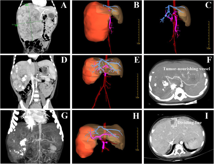
Purpose: Laparoscopic resection of giant hepatoblastoma (HB) in children has long been a subject of controversy. Here, a new procedure of two-stage laparoscopic resection of giant HB in infants was firstly reported and the feasibility was discussed.
Methods: The clinical data of three infants with HB were retrospectively reviewed, all of which received 3-5 cycles of neoadjuvant chemotherapy. Stage 1 laparoscopic selective hepatic artery ligation and liver partial partition were performed. Stage 2 laparoscopic hepatectomy was performed 2 weeks later.
Results: The results demonstrated that (1) the tumors shrank considerably in size and had relatively clear boundaries after neoadjuvant chemotherapy; (2) after stage 1 surgery, the tumor volume further reduced, while the intratumoral necrosis expanded; (3) 2 weeks later, stage 2 laparoscopic hepatectomy was performed successfully; (4) none of the cases had intraoperative complications such as tumor rupture, air embolism, hemorrhage, biliary fistula, or liver failure, and there was no recurrence or metastasis during follow-up.
Conclusions: Two-stage laparoscopic hepatectomy associating selective hepatic artery ligation and liver partial partition for HB in infants has the benefits of small invasiveness, fast recovery, improved safety, and high feasibility. However, more cases and longer follow-up are needed to assess its long-term efficacy.
Keywords: Hepatoblastoma; Infant; Laparoscopy; Staged surgery.
Conflict of interest statement
The authors declare that they have no competing interests.
Figures


Similar articles
-
Recent advances in surgical strategies and liver transplantation for hepatoblastoma.Cancer Med. 2023 Feb;12(4):3909-3918. doi: 10.1002/cam4.5300. Epub 2022 Nov 16. Cancer Med. 2023. PMID: 36394165 Free PMC article. Review.
-
Modified Associating Liver Partition and Portal Vein Ligation for Staged Hepatectomy for Hepatoblastoma in a Small Infant: How Far Can We Push the Envelope?Ann Surg. 2017 Aug;266(2):e16-e17. doi: 10.1097/SLA.0000000000002217. Ann Surg. 2017. PMID: 28288067
-
Impact of split completeness on future liver remnant hypertrophy in associating liver partition and portal vein ligation for staged hepatectomy (ALPPS) in hepatocellular carcinoma: Complete-ALPPS versus partial-ALPPS.Surgery. 2017 Feb;161(2):357-364. doi: 10.1016/j.surg.2016.07.029. Epub 2016 Sep 3. Surgery. 2017. PMID: 27596751
-
Associating liver partition with portal vein ligation and staged hepatectomy (ALPPS) for the treatment of liver tumors in children.J Pediatr Surg. 2015 Jul;50(7):1227-31. doi: 10.1016/j.jpedsurg.2014.10.019. J Pediatr Surg. 2015. PMID: 25783345
-
Associating liver partition and portal vein ligation for staged hepatectomy in patients with primary liver malignancies: A systematic review of the literature.J BUON. 2019 Jul-Aug;24(4):1371-1381. J BUON. 2019. PMID: 31646780
Cited by
-
Recent advances in surgical strategies and liver transplantation for hepatoblastoma.Cancer Med. 2023 Feb;12(4):3909-3918. doi: 10.1002/cam4.5300. Epub 2022 Nov 16. Cancer Med. 2023. PMID: 36394165 Free PMC article. Review.
-
Deploying Indocyanine Green Fluorescence-Guided Navigation System in Precise Laparoscopic Resection of Pediatric Hepatoblastoma.Cancers (Basel). 2022 Dec 9;14(24):6057. doi: 10.3390/cancers14246057. Cancers (Basel). 2022. PMID: 36551543 Free PMC article.
-
Down-Regulation of Activating Transcription Factor 3 (ATF3) in Hepatoblastoma and Its Relationship with Ferroptosis.Int J Gen Med. 2021 Dec 6;14:9401-9418. doi: 10.2147/IJGM.S340939. eCollection 2021. Int J Gen Med. 2021. PMID: 34908868 Free PMC article.
-
Case Report: ALPPS hepatectomy, an alternative to liver transplantation in central PRETEXT III hepatoblastomas: a case series.Front Pediatr. 2024 Mar 20;12:1350697. doi: 10.3389/fped.2024.1350697. eCollection 2024. Front Pediatr. 2024. PMID: 38571702 Free PMC article.
References
MeSH terms
Grants and funding
LinkOut - more resources
Full Text Sources
Other Literature Sources
Medical
