Review
. 2021 Feb 25;52(1):33.
doi: 10.1186/s13567-021-00909-x.

Intestinal organoids in farm animals

Affiliations
Review

Intestinal organoids in farm animals

Martin Beaumont et al. Vet Res. .

Abstract

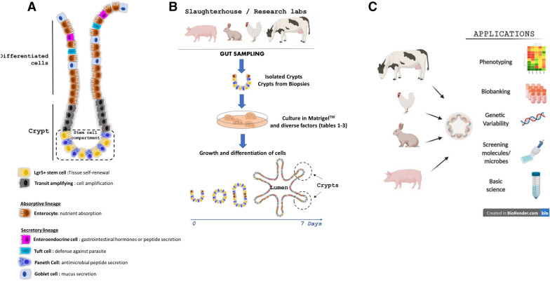
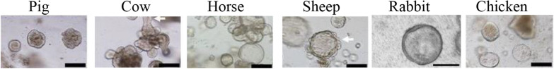
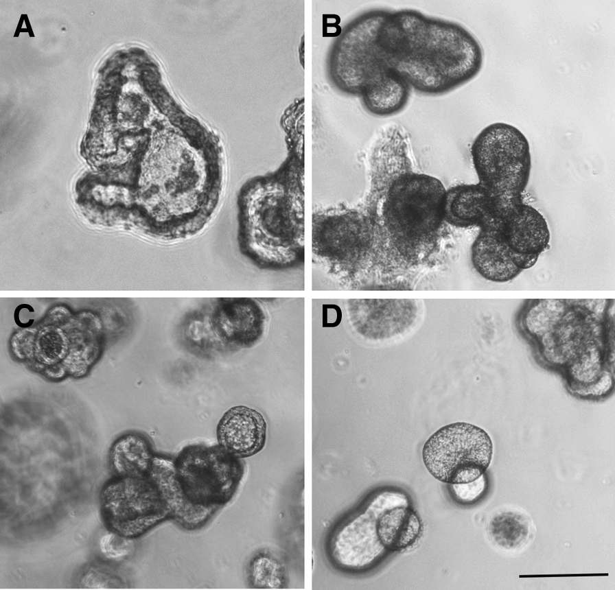
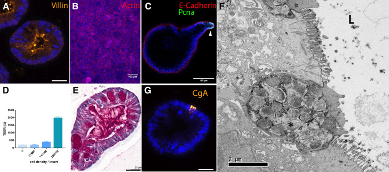
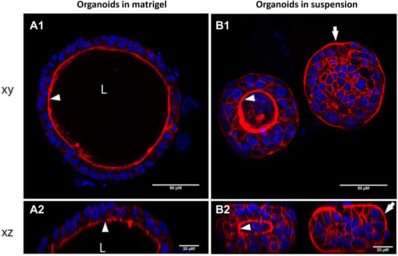
In livestock species, the monolayer of epithelial cells covering the digestive mucosa plays an essential role for nutrition and gut barrier function. However, research on farm animal intestinal epithelium has been hampered by the lack of appropriate in vitro models. Over the past decade, methods to culture livestock intestinal organoids have been developed in pig, bovine, rabbit, horse, sheep and chicken. Gut organoids from farm animals are obtained by seeding tissue-derived intestinal epithelial stem cells in a 3-dimensional culture environment reproducing in vitro the stem cell niche. These organoids can be generated rapidly within days and are formed by a monolayer of polarized epithelial cells containing the diverse differentiated epithelial progeny, recapitulating the original structure and function of the native epithelium. The phenotype of intestinal organoids is stable in long-term culture and reflects characteristics of the digestive segment of origin. Farm animal intestinal organoids can be amplified in vitro, cryopreserved and used for multiple experiments, allowing an efficient reduction of the use of live animals for experimentation. Most of the studies using livestock intestinal organoids were used to investigate host-microbe interactions at the epithelial surface, mainly focused on enteric infections with viruses, bacteria or parasites. Numerous other applications of farm animal intestinal organoids include studies on nutrient absorption, genome editing and bioactive compounds screening relevant for agricultural, veterinary and biomedical sciences. Further improvements of the methods used to culture intestinal organoids from farm animals are required to replicate more closely the intestinal tissue complexity, including the presence of non-epithelial cell types and of the gut microbiota. Harmonization of the methods used to culture livestock intestinal organoids will also be required to increase the reproducibility of the results obtained in these models. In this review, we summarize the methods used to generate and cryopreserve intestinal organoids in farm animals, present their phenotypes and discuss current and future applications of this innovative culture system of the digestive epithelium.

Keywords: Bovine; Chicken; Culture; Enteroids; Epithelium; Gut; Horse; Monolayer; Pig; Polarity; Rabbit.


Conflict of interest statement

The authors declare that they have no competing interests.

Figures

Figure 1
Figure 1
Models of intestinal organoids in farm animals. A Diverse cell lineages constitute the digestive epithelium. Paneth and stem cells are localized at the bottom of the crypts, while enterocytes, goblet cells, enteroendocrine cells and tuft cells migrate towards the lumen upon differentiation. B Schematic illustration of livestock intestinal organoid generation from epithelial crypts isolated from fresh intestinal tissue or from frozen biopsies. C Schematic representation of farm animal intestinal organoid applications in basic and applied science.
Figure 2
Figure 2
Morphology of intestinal organoids from farm animals. Brightfield images of intestinal organoids grown in Matrigel™ with 50% L-WRN conditioned media. Organoids from pig, cow, horse and sheep were obtained from the terminal ileum. Organoids from rabbit and chicken were obtained from the caecum. Scale bars: 200 µM. Images were adapted from previous publications distributed under the terms of Creative Commons Licenses: Powell and Behnke (pig, cow, horse, sheep and chicken organoids) [14] and from Mussard et al. (rabbit organoids) [16].
Figure 3
Figure 3
Morphological features of porcine gut organoids obtained from frozen tissues. Organoids are derived from: A duodenum (7 days, passage 3), B jejunum (7 days, passage 1), C ileum (7 days, passage 3) and D colon (8 days, passage 1). Observation by phase contrast microscopy. Bars: 200 µm. Images are representative of organoids obtained from 4 pigs for each digestive segment.
Figure 4
Figure 4
Characterization of intestinal organoids from farm animals. A Pig colon organoids stained for Villin (orange). Scale bar: 20 µm. B Monolayer of rabbit caecum organoid cells stained for actin (red). Scale bar: 100 µm. C Pig colon organoid stained for E-cadherin (red), and proliferating cell nuclear antigen (PCNA, green). The arrow indicates a proliferative zone in the organoid bud. Scale bar: 100 µm. D Pig colon organoid cells were seeded in Transwell inserts at several densities. Transepithelial electrical resistance (TEER) of pig organoid cell monolayers was measured 3 days post-seeding. Kruskal–Wallis test indicated a significant effect of cell density on TEER. E Pig colon organoid stained for mucins (Periodic Acid-Schiff staining). Scale bar: 20 µm. F Characterization of chicken intestinal organoid by transmission electron microscopy. The polarized organization of the cells, the brush border and the intracellular dense vesicles containing packaged mucins (marked with asterisk) were morphologically distinguishable. L: lumen. Scale bar: 2 µm. G Pig colon organoid stained for chromogranin A (CgA, orange). Scale bar: 20 µm. A, B, C and G: DNA (nuclei) is stained in blue.
Figure 5
Figure 5
Reversal of epithelial polarity in piglet colon organoid. After 7 days of culture in Matrigel™ (A), piglet colon organoids were cultured in suspension for 24 h (B). Organoids were observed by confocal laser scanning microscopy to visualize horizontal (xy, A1 and B1) and vertical (xz, A2 and B2) sections. Phalloidin staining (red) shows actin and DAPI staining (blue) shows nuclei. Arrows indicate the apical side of epithelial cells. L Lumen.
Figure 6
Figure 6
Microinjection in a pig colon organoid to access the luminal side: initial experiments using a non-toxic food dye. The micro-injection was monitored under a microscope and successively shows the insertion of the needle into the organoid (A and B), the injection of the dye into the pig organoid (B and C) and the removal of the needle (D). Organoid size: 200 µm.

Similar articles

Cited by

References

    1. Peterson LW, Artis D. Intestinal epithelial cells: regulators of barrier function and immune homeostasis. Nat Rev Immunol. 2014;14:141–153. doi: 10.1038/nri3608. - DOI - PubMed
    1. Gehart H, Clevers H. Tales from the crypt: new insights into intestinal stem cells. Nat Rev Gastroenterol Hepatol. 2019;16:19–34. doi: 10.1038/s41575-018-0081-y. - DOI - PubMed
    1. Middendorp S, Schneeberger K, Wiegerinck CL, Mokry M, Akkerman RD, van Wijngaarden S, Clevers H, Nieuwenhuis EE. Adult stem cells in the small intestine are intrinsically programmed with their location-specific function. Stem Cells. 2014;32:1083–1091. doi: 10.1002/stem.1655. - DOI - PubMed
    1. Vergauwen H (2015) The IPEC-J2 Cell Line. In: Verhoeckx K, Cotter P, Lopez-Exposito I et al. (eds) The Impact of Food Bioactives on Health: in vitro and ex vivo models. Cham (CH), pp 125–134. doi:10.1007/978-3-319-16104-4_12 - PubMed
    1. van der Hee B, Madsen O, Vervoort J, Smidt H, Wells JM. Congruence of transcription programs in adult stem cell-derived jejunum organoids and original tissue during long-term culture. Front Cell Dev Biol. 2020;8:375. doi: 10.3389/fcell.2020.00375. - DOI - PMC - PubMed

MeSH terms