. 2021 Feb 25;9(1):31.
doi: 10.1186/s40478-021-01131-8.

Trans-synaptic spreading of alpha-synuclein pathology through sensory afferents leads to sensory nerve degeneration and neuropathic pain

Affiliations

Trans-synaptic spreading of alpha-synuclein pathology through sensory afferents leads to sensory nerve degeneration and neuropathic pain

Nelson Ferreira et al. Acta Neuropathol Commun. .

Abstract

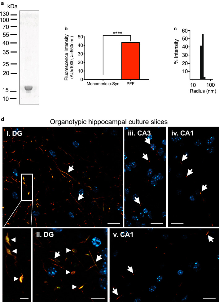
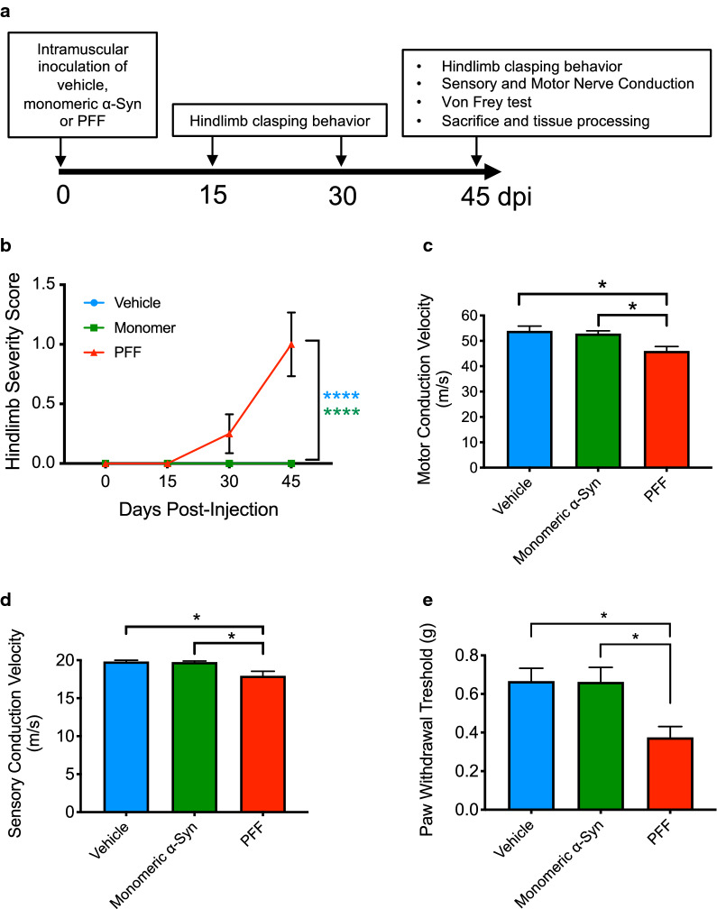
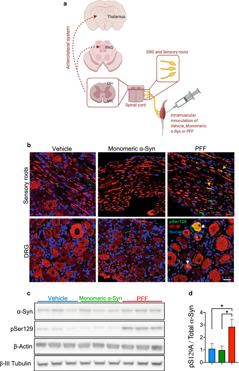
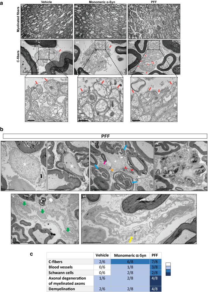
Pain is a common non-motor symptom of Parkinson's disease (PD), with current limited knowledge of its pathophysiology. Here, we show that peripheral inoculation of mouse alpha-synuclein (α-Syn) pre-formed fibrils, in a transgenic mouse model of PD, elicited retrograde trans-synaptic spreading of α-Syn pathology (pSer129) across sensory neurons and dorsal nerve roots, reaching central pain processing regions, including the spinal dorsal horn and the projections of the anterolateral system in the central nervous system (CNS). Pathological peripheral to CNS propagation of α-Syn aggregates along interconnected neuronal populations within sensory afferents, was concomitant with impaired nociceptive response, reflected by mechanical allodynia, reduced nerve conduction velocities (sensory and motor) and degeneration of small- and medium-sized myelinated fibers. Our findings show a link between the transneuronal propagation of α-Syn pathology with sensory neuron dysfunction and neuropathic impairment, suggesting promising avenues of investigation into the mechanisms underlying pain in PD.

Keywords: Alpha-synuclein; Neuropathic pain; Nociception; Parkinson’s disease; Protein aggregation.


Conflict of interest statement

The authors declare no competing interests.

Figures

Fig. 1
Fig. 1
PFF induce α-Syn aggregation and prion-like propagation in organotypic hippocampal slices. Full-length (1-140) mouse PFF were characterized using diverse biochemical and biophysical methodologies. a Coomassie blue staining after SDS-PAGE protein separation. b K114 fluorescent amyloid assay. Y-axis demonstrates the K114 fluorescence in arbitrary units. Data are shown as mean ± SEM, ****P < 0.0001 based on two-tailed unpaired t test. c Dynamic light scattering (DLS) analysis. d Following in vitro characterization and validation, PFF were injected into an organotypic hippocampal culture slice (OHCS) model. Abundant aggregation, especially at the dentate gyrus (DG), is seen, 7 days post injection (dpi) of mouse PFF, in mouse OHCS. Aggregation is observed both at the injection site (DG) and spreading throughout the hippocampal slice to the CA3 and CA1 region. Aggregates are positive for the conformation-specific MJF-14 epitope (green) and for serine-129 phosphorylation (red, antibody 11A5). Scale bars: i, iii–v: 20 µm, inset: 5 µm, ii: 10 µm
Fig. 2
Fig. 2
Pathological α-Syn induce nociceptive impairment. a Schematic illustration of the experimental design. Three-month-old M83+/− were bilaterally injected with vehicle (PBS, pH 7.4, n = 6), mouse monomeric α-Syn (2 × 5 μl at 2 mg/ml, n = 8) or mouse α-Syn PFF (2 × 5 μL at 2 mg/ml, n = 8), by intramuscular injection into the gastrocnemius muscle. Hindlimb clasping behaviour was monitored and scored regularly. At 45 dpi, mice were tested for hindlimb clasping, sensory and motor nerve conduction and plantar Von Frey filaments, before being euthanized. Tissues, including brain, spinal cord, dorsal roots and lumbar (L3–L5) dorsal root ganglia (DRG) were then snap frozen and stored at − 80 °C until use, or fixed and processed for immunohistochemistry. b Hindlimb clasping was scored on a scale from 0 to 3 as a function of dpi and displayed as mean ± SEM. The PBS- and monomeric α-Syn-injected mice did not develop clasping. Results shown as mean ± SEM, as determined by two-way ANOVA followed by Tukey’s multiple comparisons test. ****P < 0.0001. At 45 dpi mice were tested for c Motor nerve conduction velocity. Error bar indicate mean ± SEM as determined by one-way ANOVA followed by Tukey’s multiple comparison test. *P < 0.05. d Sensory nerve conduction velocity. Results shown as mean ± SEM as determined by one-way ANOVA followed by Tukey’s multiple comparison test. *P < 0.05. e Mechanical allodynia using the Von Frey test. Error bars indicate mean ± SEM as determined by one-way ANOVA followed by Tukey’s multiple comparison test. *P < 0.05
Fig. 3
Fig. 3
Peripherally injected PFF lead to prion-like α-Syn aggregation in dorsal roots and sensory neurons of the lumbar DRG. a To investigate whether the nociceptive impairment observed after intramuscular PFF-inoculation is due to the trans-synaptic propagation of α-Syn aggregates within the sensory nervous system, we performed immunodetection of PD-like α-Syn pathology in lumbar DRG and their dorsal sensory roots, spinal cord (dorsal horn and ventral horn), and brain (mesencephalic PAG and thalamic nuclei) in the vehicle, monomeric α-Syn- and PFF-injected M83+/− cohorts, at 45 dpi. Illustration was created with BioRender.com. b IHC panel showing pathological pSer129-α-Syn immunoreactivity in axons of the sensory roots (upper right panel, yellow arrows) and neuron cell bodies of the lumbar L4 DRG (lower right pane, white arrows), in PFF-injected M83 mice at 45 dpi. pSer129-α-Syn was not detected in the PBS- or monomeric α-Syn- injected cohorts. pSer129-α-Syn is represented in green, neurofilament M (NF-M) as an axonal/neuronal marker in red and nuclei are labeled in blue with Hoechst. 20 × magnification. Scale bar = 40 µm; n = 4 mice per group. c Lumbar DRG (L3–L5) homogenates from PBS- (blue, n = 6), monomeric- (green, n = 6) and PFF-injected (red, n = 5) were separated by SDS-PAGE and visualized by immunoblotting with anti-α-Syn antibody, anti-pSer129-α-Syn, anti-β-actin, and anti-β-III tubulin. d Densitometry quantification of immunoblots as a ratio of pSer129/total α-Syn. Results shown as mean ± SEM as determined by one-way ANOVA followed by Tukey’s multiple comparison test. *P < 0.05
Fig. 4
Fig. 4
Intramuscular injection of α-Syn PFF prompt pathological aggregation of α-Syn in neurons of the lumbar spinal cord, midbrain periaqueductal grey and thalamus. a Panoramic views: pSer129-α-Syn (in green) co-detection with neuronal nuclei antigen (NeuN, in red) in vehicle- (PBS, pH 7.4, left panel) and PFF-injected mice (right panel) lumbar spinal cord sections at 45 dpi (DH, dorsal horn; VH, ventral horn), b pSer129-α-Syn (in green) co-detection with neuronal nuclei antigen (NeuN, in red) in α-Syn PFF- injected mice. c Astrogliosis in the lumbar spinal cord, midbrain periaqueductal grey and thalamus was observed in the PFF-injected cohort. pSer129-α-Syn (in green) and glial fibrillary acidic protein marker (GFAP, in red) immunoreactivity, VH and DH of lumbar spinal cord, d midbrain periaqueductual grey (MB-PAG) and e thalamus. DAPI (blue) was used to stain the nuclei. Scale bar = 100 µm; insets in merge show 63X magnified views
Fig. 5
Fig. 5
Peripherally injected PFF induces small- and medium-sized myelinated fiber pathology in the dorsal roots. a Morphological evaluation of the dorsal sensory roots by EM. Representative low magnification images of the sensory roots from vehicle- (panel i, PBS, pH 7.4), monomeric α-Syn (panel ii) and α-Syn PFF-injected mice (panel iii), denoting normal distribution of myelinated fibers with some morphological abnormalities identified in the PFF cohort (panel iii, white arrows). Scale bar 20 μm. Panels iv–ix denote high magnification pictures (scale bar = 2 µm), and respective insets (scale bar = 250 nm), showing an abnormal structure of the Remak bundles composed of numerous unmyelinated axons enclosed by basement membrane of Schwann cells, particularly in the monomeric and PFF- injected mice (panel vi–ix). Note electron loose Schwann cell processes, and the lack of these processes around some of the axons (orange arrows). b Image displaying completely (panel i) or partially demyelinated axon (panel ii) (> 1.5 μm diameter; white asterisks). In Panel (i), flat sheets of Schwann cell cytoplasmic processes enclosed by a common basement membrane, designated as bands of Büngner (black arrow) that are usually formed after degeneration of unmyelinated axons, can also be observed. Panel (iii) Picture showing degeneration of small- and medium-sized myelinated fibers (blue arrows), Schwann cell cytoplasm containing degradation products and myelin ovoids (red arrowhead), free debris in the endoneurium (orange arrow) and disorganized Remak bundles with few unmyelinated C-fibers (pink arrow). Panel (iv) A macrophage engulfing myelin fragments is highlighted. Panel (v) Green arrows feature projection of endothelial cell nuclei into the lumen of blood vessels. Panel (vi) Exhibits an endothelial cell with abnormal morphology, with irregular small branches and swollen electron loose cytoplasm (yellow arrow). Scale bars are 2 μm (Panels i, vi) and 5 μm (Panels ii, iii, iv, v). c Dorsal sensory roots semi-quantitative pathology scoring summary of the three cohorts in this study; the number of mice with detectable pathological abnormalities in C-fibers, blood vessels, Schwan cells or axonal degeneration and demyelination in the endoneurium (n/6 for vehicle- injected mice and n/8 for both monomeric α-Syn- and PFF-injected mice) is shown along with density of pathology represented by colour where darker colour indicates aggravated pathology

Similar articles

Cited by

References

    1. Abdelmotilib H, Maltbie T, Delic V, Liu Z, Hu X, Fraser KB, Moehle MS, Stoyka L, Anabtawi N, Krendelchtchikova V, et al. alpha-Synuclein fibril-induced inclusion spread in rats and mice correlates with dopaminergic Neurodegeneration. Neurobiol Dis. 2017;105:84–98. doi: 10.1016/j.nbd.2017.05.014. - DOI - PMC - PubMed
    1. Arancibia-Carcamo IL, Ford MC, Cossell L, Ishida K, Tohyama K, Attwell D. Node of Ranvier length as a potential regulator of myelinated axon conduction speed. Elife. 2017;6:e23329. doi: 10.7554/eLife.23329. - DOI - PMC - PubMed
    1. Ayers JI, Riffe CJ, Sorrentino ZA, Diamond J, Fagerli E, Brooks M, Galaleldeen A, Hart PJ, Giasson BI. Localized induction of wild-type and mutant alpha-synuclein aggregation reveals propagation along neuroanatomical tracts. J Virol. 2018 doi: 10.1128/JVI.00586-18. - DOI - PMC - PubMed
    1. Bala U, Leong MP, Lim CL, Shahar HK, Othman F, Lai MI, Law ZK, Ramli K, Htwe O, Ling KH, et al. Defects in nerve conduction velocity and different muscle fibre-type specificity contribute to muscle weakness in Ts1Cje Down syndrome mouse model. PLoS ONE. 2018;13:e0197711. doi: 10.1371/journal.pone.0197711. - DOI - PMC - PubMed
    1. Beiske AG, Loge JH, Ronningen A, Svensson E. Pain in Parkinson's disease: prevalence and characteristics. Pain. 2009;141:173–177. doi: 10.1016/j.pain.2008.12.004. - DOI - PubMed

Publication types

MeSH terms