

Idiopathic superficial siderosis of the central nervous system
- PMID: 33632336
- PMCID: PMC7908722
- DOI: 10.1186/s40673-021-00133-5
Idiopathic superficial siderosis of the central nervous system
Abstract
Background: Regardless of the cause of the superficial siderosis (SS) disease, which is bleeding, the source of bleeding cannot be found in some cases.
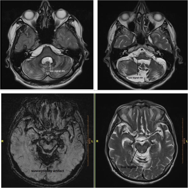
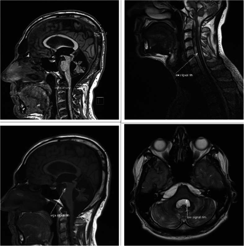
Case presentation: In this article, we report two cases with idiopathic SS. Case 1 presented with bilateral hearing loss, cognitive impairment, sleep disturbances, and tremors. Case 2 presented with sensory neural hearing loss, ataxia, and spastic paraparesis. In both cases, brain MRI indicated evidence of SS. CT myelogram and SPECT with labeled RBC couldn't help finding the source of occult bleeding.
Conclusion: SS is a rare central nervous system disease caused by the deposition of hemosiderin in the brain and spinal cord, which results in the progression of neurological deficits. The cause of this hemorrhage is often subarachnoid haemorrhage, intracranial surgery, carcinoma, arteriovenous malformation, nerve root avulsion, and dural abnormality. The condition progresses slowly and, by the time diagnosis is confirmed, the damage is often irreversible. In our cases, brain MRI clarified the definitive diagnosis, but we could not find the source of bleeding. SS should be considered in cases with ataxia and hearing loss, even if no source of bleeding is found.
Keywords: Chronic subarachnoid hemorrhage; Hemosiderin; Sensorineural deafness; Superficial siderosis.
Conflict of interest statement
There is no competing interest to report.
Figures
Similar articles
-
Clival meningocele causing bilateral hearing loss in a child due to superficial siderosis of the central nervous system: case report.J Neurosurg Pediatr. 2018 May;21(5):498-503. doi: 10.3171/2017.11.PEDS17302. Epub 2018 Feb 16. J Neurosurg Pediatr. 2018. PMID: 29451456
-
Superficial siderosis of the central nervous system associated with ventral dural defects: bleeding from the epidural venous plexus.J Neurol. 2021 Apr;268(4):1491-1494. doi: 10.1007/s00415-020-10319-2. Epub 2021 Jan 3. J Neurol. 2021. PMID: 33389031
-
Superficial siderosis.Neurology. 2006 Apr 25;66(8):1144-52. doi: 10.1212/01.wnl.0000208510.76323.5b. Neurology. 2006. PMID: 16636229
-
[Superficial siderosis: case report and literature review].Srp Arh Celok Lek. 2013 Mar-Apr;141(3-4):219-22. doi: 10.2298/sarh1304219k. Srp Arh Celok Lek. 2013. PMID: 23745347 Review. Serbian.
-
Dural closure for the treatment of superficial siderosis.J Neurosurg Spine. 2013 Apr;18(4):388-93. doi: 10.3171/2013.1.SPINE12649. Epub 2013 Feb 22. J Neurosurg Spine. 2013. PMID: 23432322 Review.
Cited by
-
Superficial siderosis: comparison of two cases indicates two distinct diagnostic entities. Illustrative cases.J Neurosurg Case Lessons. 2023 May 29;5(22):CASE23161. doi: 10.3171/CASE23161. Print 2023 May 29. J Neurosurg Case Lessons. 2023. PMID: 37249139 Free PMC article.
-
A case report of superficial siderosis of the central nervous system and literature review.J Int Med Res. 2023 Sep;51(9):3000605231198389. doi: 10.1177/03000605231198389. J Int Med Res. 2023. PMID: 37702555 Free PMC article. Review.
-
Ataxias: Hereditary, Acquired, and Reversible Etiologies.Semin Neurol. 2023 Feb;43(1):48-64. doi: 10.1055/s-0043-1763511. Epub 2023 Feb 24. Semin Neurol. 2023. PMID: 36828010 Free PMC article.
References
LinkOut - more resources
Full Text Sources
Other Literature Sources
