. 2021:177:345-357.
doi: 10.1016/B978-0-12-819814-8.00021-4.

The role of biomarkers and neuroimaging in ischemic/hemorrhagic risk assessment for cardiovascular/cerebrovascular disease prevention

Affiliations

The role of biomarkers and neuroimaging in ischemic/hemorrhagic risk assessment for cardiovascular/cerebrovascular disease prevention

Elif Gokcal et al. Handb Clin Neurol. 2021.

Abstract

Stroke prevention in patients with atrial fibrillation is arguably one of the fastest developing areas in preventive medicine. The increasing use of direct oral anticoagulants and nonpharmacologic methods such as left atrial appendage closure for stroke prevention in these patients has increased clinicians' options for optimal care. Platelet antiaggregants are also commonly used in other ischemic cardiovascular and or cerebrovascular conditions. Long term use of oral anticoagulants for atrial fibrillation is associated with elevated risks of major bleeds including especially brain hemorrhages, which are known to have extremely poor outcomes. Neuroimaging and other biomarkers have been validated to stratify brain hemorrhage risk among older adults. A thorough understanding of these biomarkers is essential for selection of appropriate anticoagulant or left atrial appendage closure for stroke prevention in patients with atrial fibrillation. This article will address advances in the stratification of ischemic and hemorrhagic stroke risk among patients with atrial fibrillation and other conditions.

Keywords: Anticoagulation; Antithrombotic; Cerebral amyloid angiopathy; Hypertensive cerebral hemorrhage; Intracerebral hemorrhage; Platelet antiaggregants; Risk scores; Stroke prevention.


Figures

Fig. 30.1.
Fig. 30.1.
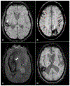
Hemorrhagic imaging markers in different types of cerebral small vessel diseases. (A) Patient with CAA with a lobar ICH (arrow, right temporal) and strictly lobar CMBs (stars). (B) Patient with CAA with a lobar ICH (arrow, left occipital), strictly lobar CMBs (stars), and multifocal cSS (circle/ellipses). (C) Patient with HTN-cSVD with a deep ICH (arrow, right basal ganglia) and a CMB located in deep brain region (star, left thalamus). (D) Patient with mixed ICH/CMB with a lobar ICH (arrow, right parietotemporal), lobar CMB (star, left frontal) as well as deeply located CMBs (stars, right and left thalamus). All MRI images are SWI-MRI sequences. The arrow represents ICH, the star represents CMB, and the oval shape represents cSS. CAA, cerebral amyloid angiopathy; CMB, cerebral microbleeds; cSS, cortical superficial siderosis; HTN-cSVD, hypertensive cerebral small vessel disease; ICH, intracerebral hemorrhage; SWI-MRI, susceptibility-weighted imaging-magnetic resonance imaging sequence.
Fig. 30.2.
Fig. 30.2.
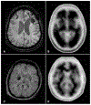
Florbetapir PET for diagnosis of cerebral amyloid angiopathy. (A–B) represent a patient who had a left frontal intracerebral hemorrhage (ICH) and right frontal cortical superficial siderosis on susceptibility-weighted imaging (SWI, Panel A), fulfilling neuroradiological criteria for cerebral amyloid angiopathy (CAA). Florbetapir PET (Panel B) in this CAA patient shows loss of contrast between the cortex and subcortical white matter (positive scan), indicating intense Florbetapir uptake in the cortex because of high vascular amyloid load. The patient in (C–D) had a hypertensive deep right basal ganglia hemorrhage visible on SWI MRI (Panel C). In this patient’s Florbetapir PET (Panel D), the contrast between cortex and subcortical structures is preserved (negative scan), confirming low amyloid load in the cortex. Modified from Gurol ME, Becker JA, Fotiadis P, et al. (2016). Florbetapir-PET to diagnose cerebral amyloid angiopathy: A prospective study. Neurology 87: 2043–2049 with permission. Copyright ©2016, Wolters Kluwer Health, Inc.
Fig. 30.3.
Fig. 30.3.
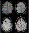
Representative examples of the cortical superficial siderosis (cSS) multifocality score. To assess multifocality of cSS, each hemisphere is scored separately for cSS, as following: 0: none; 1: 1–3 immediately adjacent sulci with cSS; and 2: >3 immediately adjacent sulci (disseminated) or >1 nonadjacent sulci with cSS. The total score is derived by adding the right and left hemisphere scores (range 0–4). Examples are presented on the figure, and the score for each hemisphere (0,1,2) is provided in bottom corners of each patient scan. (A) Patient with a total cSS score of 1 (1 for right and 0 for left hemisphere). (B) Patient with a total cSS score of 2 (0 for right and 2 for left hemisphere). (C) Patient with a total cSS score of 3 (1 for right and 2 for left hemisphere). (D) Patient with a total cSS score of 4 (2 for right and 2 for left hemisphere). All MRI images are SWI-MRI sequences. SWI-MRI, susceptibility-weighted imaging-magnetic resonance imaging.
Fig. 30.4.
Fig. 30.4.
White matter hyperintensities (WMH) on fluid attenuated inversion recovery (FLAIR) MRI sequence. (A) Mild FLAIR white matter hyperintensities (WMH), (B) moderate WMH, and (C) severe WMH.

Similar articles

Cited by

References

    1. Baczek VL, Chen WT, Kluger J et al. (2012). Predictors of warfarin use in atrial fibrillation in the United States: a systematic review and meta-analysis. BMC Fam Pract 13: 5. - PMC - PubMed
    1. Bekwelem W, Connolly SJ, Halperin JL et al. (2015). Extracranial systemic embolic events in patients with nonvalvular atrial fibrillation: incidence, risk factors, and outcomes. Circulation 132: 796–803. - PubMed
    1. Benjamin EJ, Blaha MJ, Chiuve SE et al. (2017). Heart disease and stroke statistics-2017 update: a report from the American Heart Association. Circulation 135: e146–e603. - PMC - PubMed
    1. Blackshear JL, Odell JA (1996). Appendage obliteration to reduce stroke in cardiac surgical patients with atrial fibrillation. Ann Thorac Surg 61: 755–759. - PubMed
    1. Boersma LV, Schmidt B, Betts TR et al. (2016). Implant success and safety of left atrial appendage closure with the WATCHMAN device: peri-procedural outcomes from the EWOLUTION registry. Eur Heart J 37: 2465–2474. - PMC - PubMed

LinkOut - more resources