. 2021 Mar 2;77(8):1073-1088.
doi: 10.1016/j.jacc.2020.12.060.

Mitochondria-Rich Extracellular Vesicles From Autologous Stem Cell-Derived Cardiomyocytes Restore Energetics of Ischemic Myocardium

Affiliations

Mitochondria-Rich Extracellular Vesicles From Autologous Stem Cell-Derived Cardiomyocytes Restore Energetics of Ischemic Myocardium

Gentaro Ikeda et al. J Am Coll Cardiol. .

Abstract

Background: Mitochondrial dysfunction results in an imbalance between energy supply and demand in a failing heart. An innovative therapy that targets the intracellular bioenergetics directly through mitochondria transfer may be necessary.

Objectives: The purpose of this study was to establish a preclinical proof-of-concept that extracellular vesicle (EV)-mediated transfer of autologous mitochondria and their related energy source enhance cardiac function through restoration of myocardial bioenergetics.

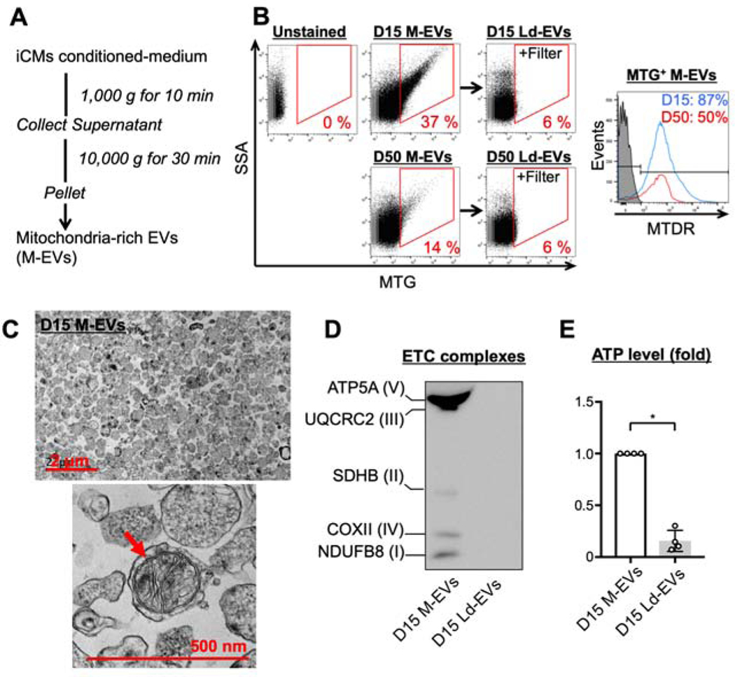
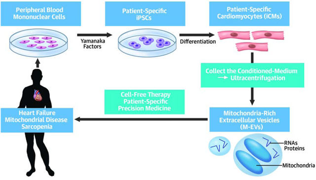
Methods: Human-induced pluripotent stem cell-derived cardiomyocytes (iCMs) were employed. iCM-conditioned medium was ultracentrifuged to collect mitochondria-rich EVs (M-EVs). Therapeutic effects of M-EVs were investigated using in vivo murine myocardial infarction (MI) model.

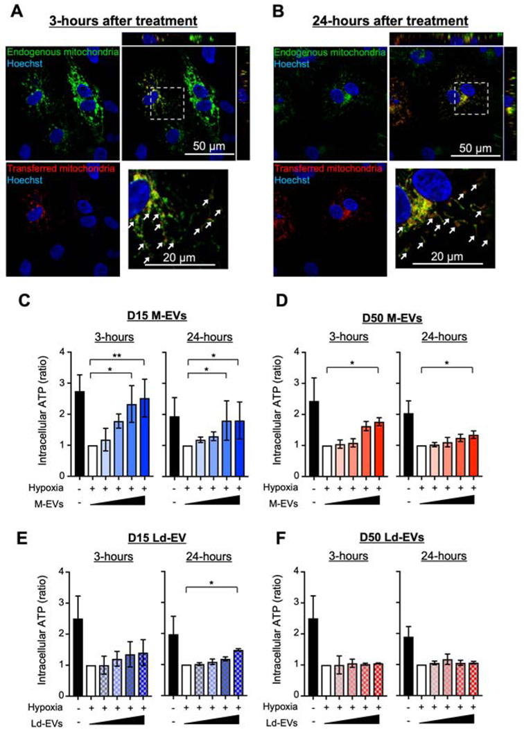
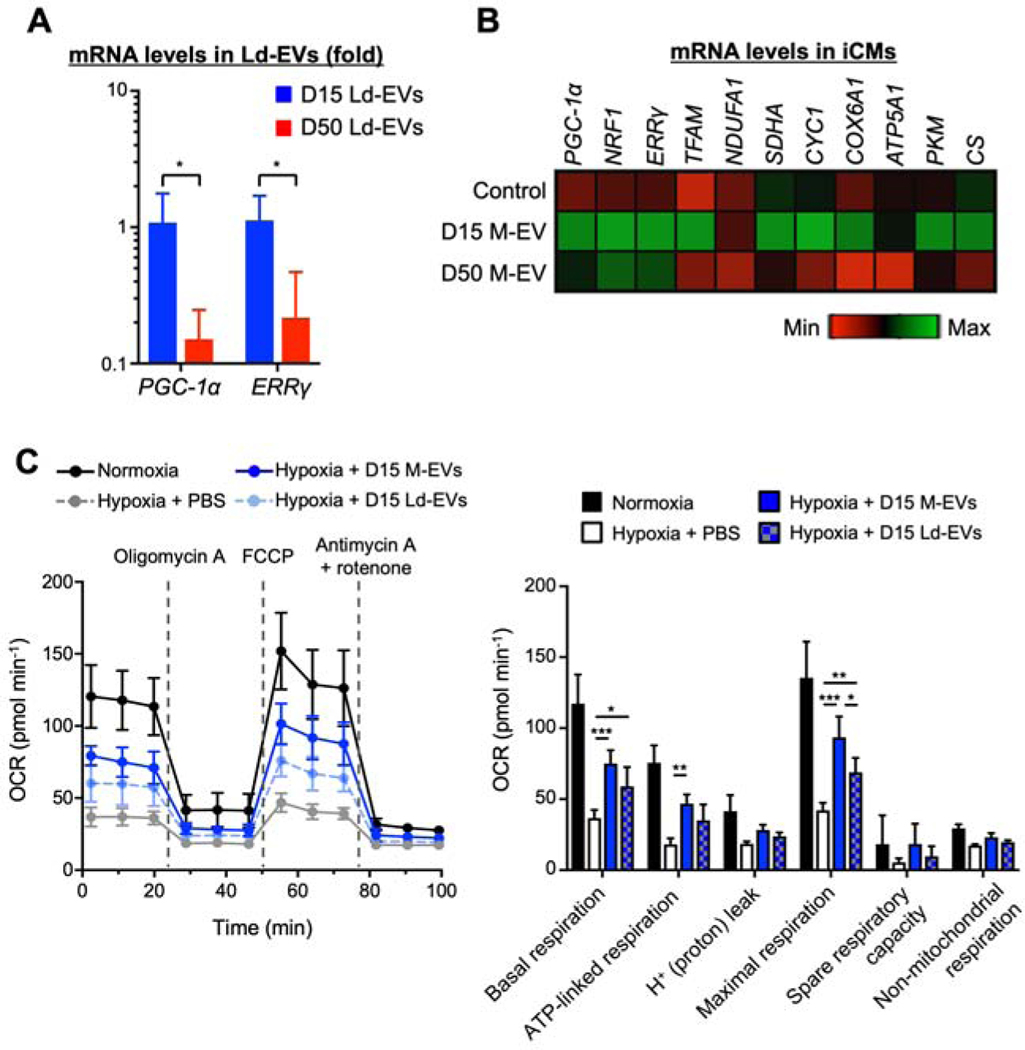
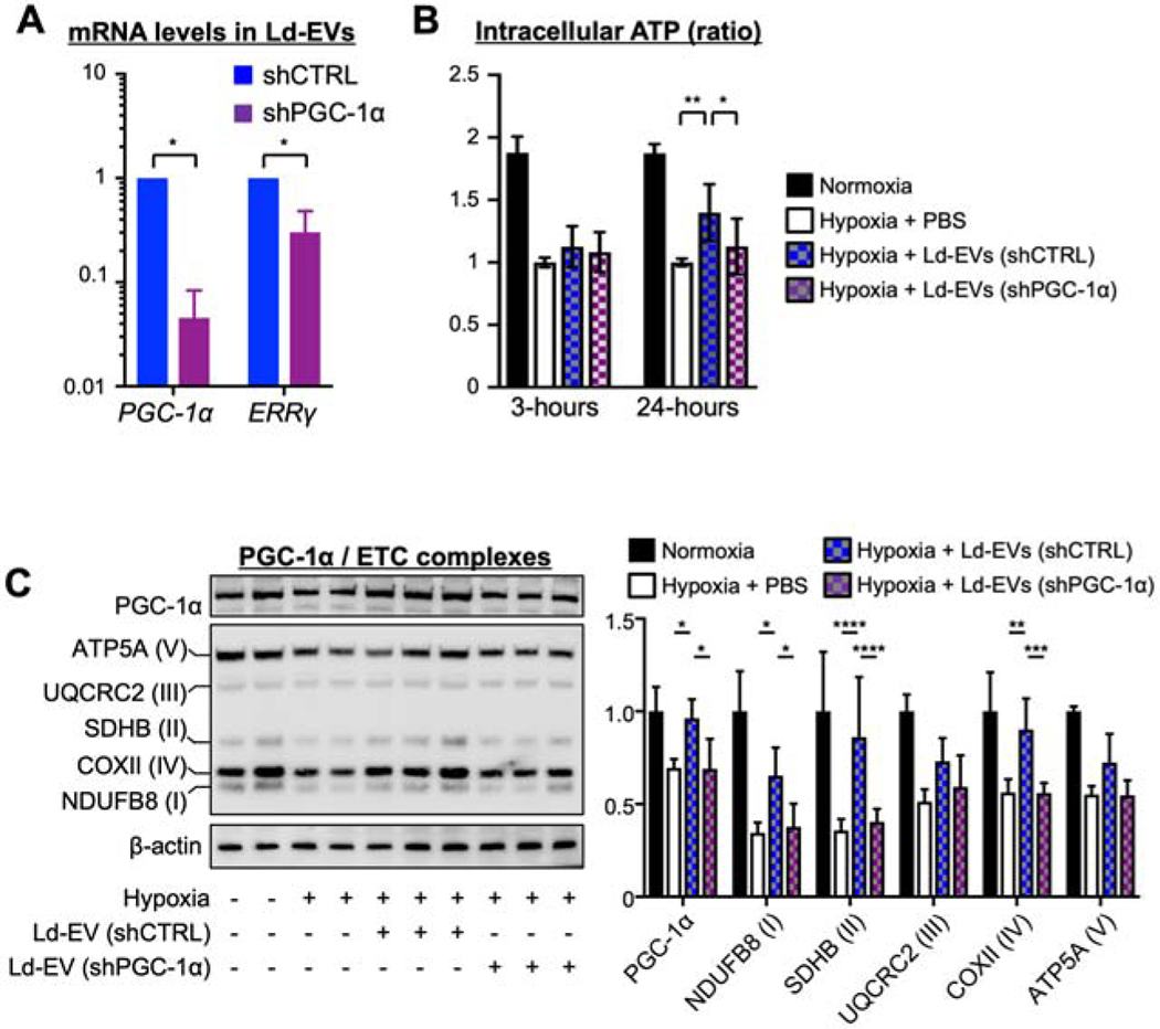
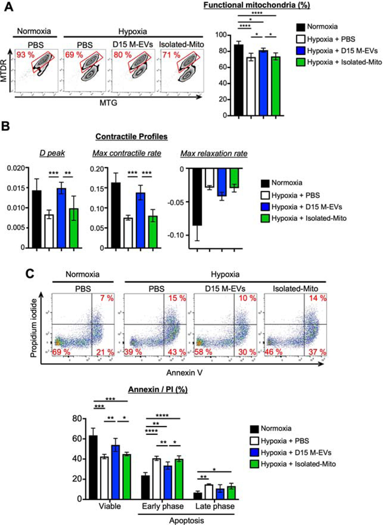
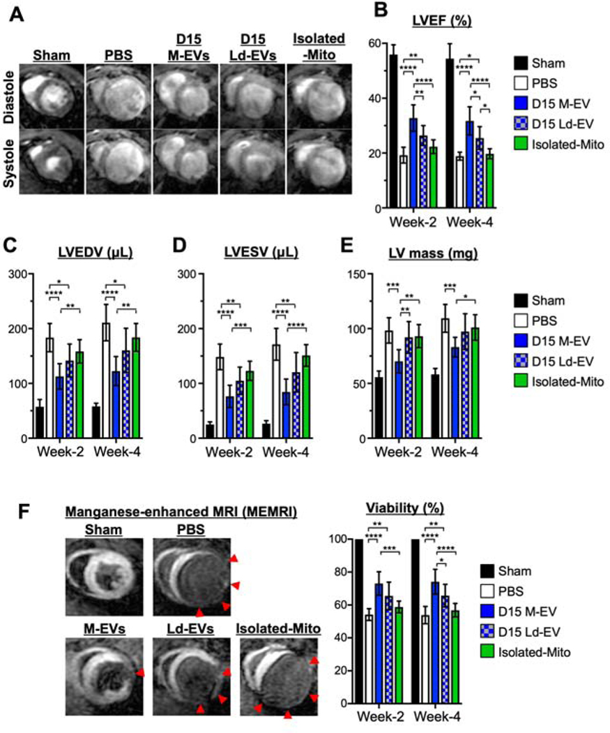
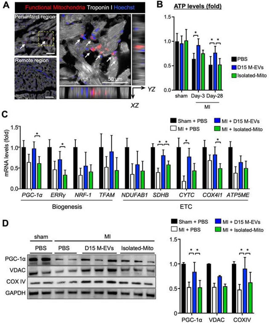
Results: Electron microscopy revealed healthy-shaped mitochondria inside M-EVs. Confocal microscopy showed that M-EV-derived mitochondria were transferred into the recipient iCMs and fused with their endogenous mitochondrial networks. Treatment with 1.0 × 10⁸/ml M-EVs significantly restored the intracellular adenosine triphosphate production and improved contractile profiles of hypoxia-injured iCMs as early as 3 h after treatment. In contrast, isolated mitochondria that contained 300× more mitochondrial proteins than 1.0 × 10⁸/ml M-EVs showed no effect after 24 h. M-EVs contained mitochondrial biogenesis-related messenger ribonucleic acids, including proliferator-activated receptor γ coactivator-1α, which on transfer activated mitochondrial biogenesis in the recipient iCMs at 24 h after treatment. Finally, intramyocardial injection of 1.0 × 10⁸ M-EVs demonstrated significantly improved post-MI cardiac function through restoration of bioenergetics and mitochondrial biogenesis.

Conclusions: M-EVs facilitated immediate transfer of their mitochondrial and nonmitochondrial cargos, contributing to improved intracellular energetics in vitro. Intramyocardial injection of M-EVs enhanced post-MI cardiac function in vivo. This therapy can be developed as a novel, precision therapeutic for mitochondria-related diseases including heart failure.

Keywords: bioenergetics; heart failure; human stem cells; mitochondria; myocardial infarction.


Conflict of interest statement

Funding Support and Author Disclosures Dr. Ikeda has received funding support through the Stanford Dean’s Postdoctoral Fellowship, Japan Heart Foundation/Bayer Yakuhin, and an American Heart Association postdoctoral fellowship. Ms. Santoso has received funding support through the Alpha Omega Alpha Carolyn B. Kuckein Student Research Fellowship. Dr. Yang has received funding support through National Institutes of Health, National Heart, Lung, and Blood Institute grants 1 K24 HL130553K and UM1 L12026; and funding support from SPARK Stanford University funding support through Stanford Cardiovascular Institute Seed Grant. All other authors have reported that they have no relationships relevant to the contents of this paper to disclose.

Figures

Figure 1.
Figure 1.. Characterization of mitochondria-rich extracellular vesicles (M-EVs).
A. Differential ultracentrifugation was employed to isolate M-EVs. B. Representative flow cytometry (FCM) dot plots and histograms of M-EVs and Large vesicle-depleted EVs (Ld-EVs). C. Transmission electron microscopy of D15 M-EVs. D. The protein expression of the electron transport chain (ETC) in D15 M-EVs. E. M-EVs, but not Ld-EVs, produced extracellular ATP (n=4/group). *P<0.0001 by an unpaired t-test. ATP5A=ATP synthase α-subunit; COX II=cytochrome c oxidase subunit-2; MTDR=mitotracker deep red; MTG=mitotracker green; MTT=3-(4,5-dimethylthiazol-2-yl)-2,5-diphenyltetrazolium bromide; NDUFB8=NADH dehydrogenase-1β subcomplex subunit-8; SDHB=succinate dehydrogenase complex iron sulfur subunit B; SSA=side-scatter-area; UQCRC2=ubiquinol-cytochrome c reductase core protein-2.
Figure 2.
Figure 2.. M-EVs facilitate transfer of their mitochondrial cargo and restore intracellular bioenergetics in hypoxia-injured iCMs.
A and B. Representative microscopic image of iCMs treated with M-EVs containing RFP+-mitochondria. Transferred M-EV-mitochondria (red) were observed in the recipient iCMs harboring GFP+-mitochondria (green). Some of the transferred mitochondria were fused with the native mitochondrial networks (white arrows). Scale bar: 50 and 20 μm. C to F. ATP levels in iCMs treated with M-EVs or Ld-EVs. (0, 1.0×10⁷, 3.0×10⁷, 1.0×10⁸ or 3.0×10⁸/mL: wedge). Left, 3-hours after treatment. Right, 24-hours after treatment (n=4–6/group). *P<0.05 and **P<0.001 by a one-way ANOVA followed by Tukey’s test.
Figure 3.
Figure 3.. Non-mitochondrial cargo in D15 M-EVs restored bioenergetics through activation of PGC-1α-mediated mitochondrial biogenesis in the recipient iCMs.
A. PGC-1α and ERRγ mRNA, normalized to U6, were upregulated in D15 Ld-EVs (n=4/group). *P<0.05 by an unpaired t-test. B. Quantitative gene expression in iCMs, normalized to ACTB shown as fold change relative to normoxic iCMs (n=4–5/group). C. Seahorse extracellular-flux assays measuring oxygen-consumption rate (OCR) (n=5–6/group). *P<0.01, **P<0.001 and ***P<0.0001 by a one-way ANOVA followed by Tukey’s test. ATP5A1=ATP synthase F1 α-subunit; COX6A1=cytochrome c oxidase subunit-6A1; Cs=citrate synthase; CYC1=cytochrome c1; ERRγ=estrogen-related receptor γ; FCCP=carbonyl cyanide-4-(trifluoromethoxy)phenylhydrazone; NRF1=Nuclear respiratory factor-1; NDUFA1=NADH dehydrogenase-1α subcomplex subunit-1; PGC-1α=peroxisome proliferator-activated receptor γ coactivator 1α; PKM=pyruvate kinase M1/2; SDHA=succinate dehydrogenase complex flavoprotein subunit-A; TFAM=mitochondrial transcription factor-A.
Figure 4.
Figure 4.. PGC-1α knockdown compromised the restoration of mitochondrial bioenergetics and biogenesis in the hypoxia-injured iCMs.
A. Relative PGC-1α and ERRγ mRNA expression, normalized to U6, in Ld-EVs from D15 transduced with pLKO.1 expressing control shRNA (shCTRL) or anti-PGC-1α shRNA (shPGC-1α) (n=3/group). *P<0.0001 by an unpaired t-test. B. PGC-1α knockdown reduced restoration of ATP levels in hypoxia-injured iCMs at 24hous (n=5–8/group). C. Representative immunoblots and quantitative analyses of PGC-1α, ETC complexes and β-actin (n=4–6/group). *P<0.05, **P<0.01, ***P<0.001 and ****P<0.0001 by a one-way ANOVA followed by Tukey’s test. Abbreviations as in Figure 1.
Figure 5.
Figure 5.. D15 M-EVs improved mitochondrial function, contractile property, and cell survival in hypoxia-injured iCMs.
A. Representative FCM dotplots of iCMs labeled with MTG/MTDR (n=5–6/group). B. D peak and maximum contractile rate in iCMs (n=5–6/group). C. Representative FCM dotplots of iCMs stained with Annexin V/Propidium iodide (n=4–5/group). *P<0.05, **P<0.01, ***P<0.001 and ****P<0.0001 by a one-way ANOVA followed by Tukey’s test.
Figure 6.
Figure 6.. The effects of intramyocardial injection of D15 M-EVs on post-myocardial infarction (MI) cardiac remodeling.
A. Representative images of short-axis acquisitions at mid-left ventricle (LV). B to E. LV ejection fraction (LVEF), LV end-diastolic volume (LVEDV), LV end-systolic volume (LVESV) and LV mass at weeks-2 and −4 after MI, evaluated by MRI. F. Representative images and quantitative analyses of myocardial viability as visualized by manganese-enhanced MRI (MEMRI). Red arrows: non-viable regions (n=8/group). *P<0.05, **P<0.01, ***P<0.001 and ****P<0.0001 by a two-way ANOVA followed by Tukey’s test.
Figure 7.
Figure 7.. D15 M-EV therapy enhances bioenergetics and PGC-1α-mediated mitochondrial biogenesis in the peri-infarct region.
A. MTDR+-mitochondria were detected inside the Troponin I+-cardiomyocytes, highlighted with white arrows. Scale bar: 50 μm. B. Tissue ATP levels, normalized by tissue protein levels (n=4–6/group). C. Mitochondrial biogenesis-related gene expression levels in the peri-infarct region, normalized to GAPDH, measured by RT-PCR (n=4–5/group). D. Representative immunoblots and quantitative analyses of PGC-1α, VDAC and COX IV, normalized to GAPDH (n=4–5/group). *P<0.05 by a one-way ANOVA followed by Tukey’s test. ATP5ME=ATP synthase subunit-e; COX IV=cytochrome c oxidase subunit-4; COX4I1=cytochrome c oxidase subunit-4 isoform-1; NDUFAB1=NADH ubiquinone oxidoreductase subunit-AB1; VDAC=voltage-dependent anion channels; other abbreviations as in Figure 3.
Central Illustration.
Central Illustration.. Extracellular Vesicle-Mediated Autologous Mitochondrial Transplantation.
We obtained human-induced pluripotent stem cells (iPSCs)-derived cardiomyocytes (iCMs) and succeeded in collecting mitochondria-rich extracellular vesicles (M-EVs) from iCM conditioned-medium. The autologous M-EVs can be applied to patients with mitochondria-related diseases including heart failure.

Comment in

Similar articles

Cited by

References

    1. Yancy CW, Jessup M, Bozkurt B, et al. 2013 ACCF/AHA guideline for the management of heart failure: executive summary: a report of the American College of Cardiology Foundation/American Heart Association Task Force on practice guidelines. J Am Coll Cardiol. 2013. October 15;62(16):e147–239. - PubMed
    1. Taylor CJ, Ordóñez-Mena JM, Roalfe AK, et al. Trends in survival after a diagnosis of heart failure in the United Kingdom 2000–2017:population based cohort study. BMJ. 2019;364:1223. - PMC - PubMed
    1. Neubauer S. The failing heart-an engine out of fuel. N Engl J Med. 2007;356:1140–1151. - PubMed
    1. Sekiguchi M. Electron microscopical observations of the myocardium in patients with idiopathic cardiomyopathy using endomyocardial biopsy. J Mol Cell Cardiol. 1974;6:111–122. - PubMed
    1. Starling RC, Hammer DF, Altschuld RA. Human myocardial ATP content and in vivo contractile function. Mol Cell Biochem. 1998;180:171–177. - PubMed

MeSH terms

Substances