. 2021 May;42(5):975-979.
doi: 10.3174/ajnr.A7019. Epub 2021 Feb 25.

Widening the Neuroimaging Features of Adenosine Deaminase 2 Deficiency

Affiliations

Widening the Neuroimaging Features of Adenosine Deaminase 2 Deficiency

A F Geraldo et al. AJNR Am J Neuroradiol. 2021 May.

Abstract

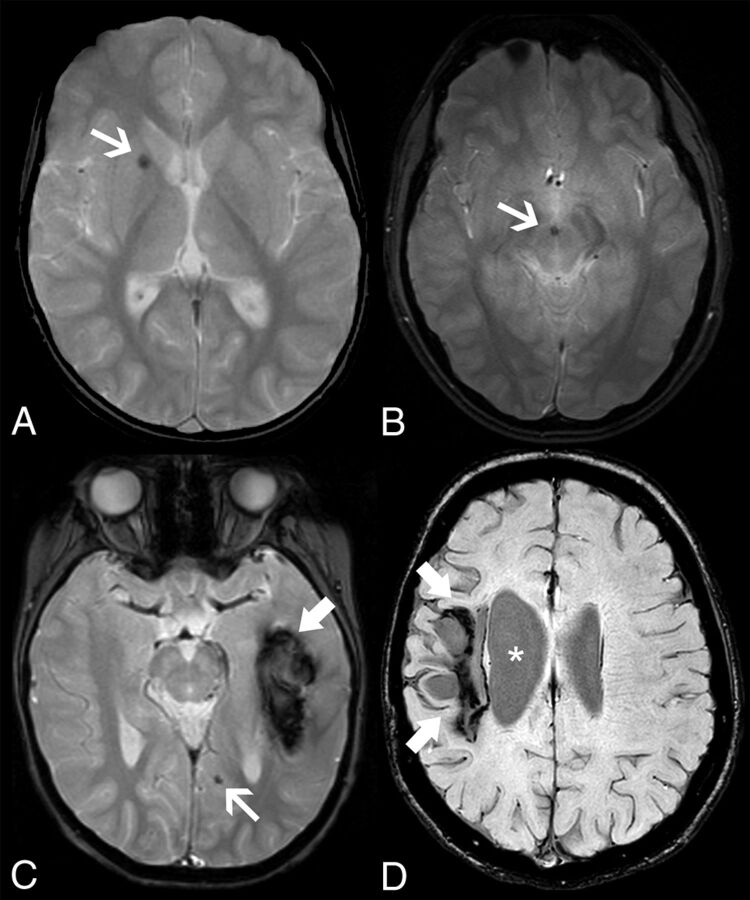
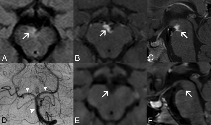
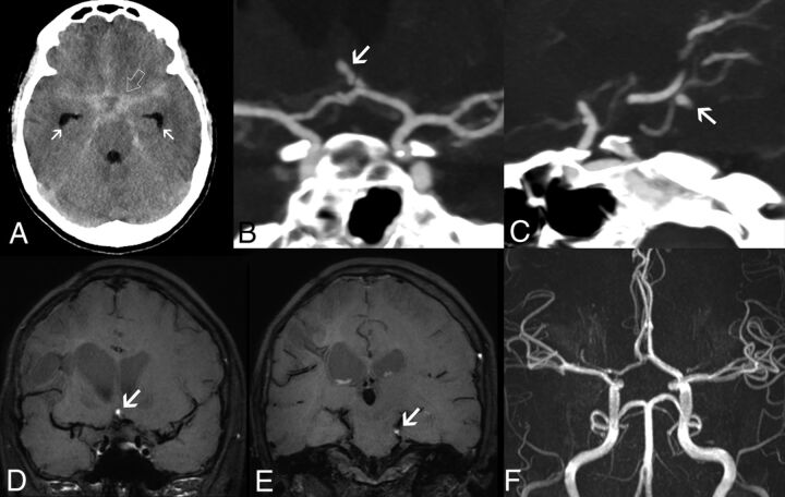
Adenosine deaminase 2 deficiency (OMIM #615688) is an autosomal recessive disorder characterized by a wide clinical spectrum, including small- and medium-sized vessel vasculopathies, but data focusing on the associated neuroimaging features are still scarce in the literature. Here, we describe the clinical neuroimaging features of 12 patients with genetically proven adenosine deaminase 2 deficiency (6 males; median age at disease onset, 1.3 years; median age at genetic diagnosis, 15.5 years). Our findings expand the neuroimaging phenotype of this condition demonstrating, in addition to multiple, recurrent brain lacunar ischemic and/or hemorrhagic strokes, spinal infarcts, and intracranial aneurysms, also cerebral microbleeds and a peculiar, likely inflammatory, perivascular tissue in the basal and peripontine cisterns. Together with early clinical onset, positive family history, inflammatory flares and systemic abnormalities, these findings should raise the suspicion of adenosine deaminase 2 deficiency, thus prompting genetic evaluation and institution of tumor necrosis factor inhibitors, with a potential great impact on neurologic outcome.


Figures

FIG 1.
FIG 1.
Intracranial hemorrhagic manifestations in adenosine deaminase 2 deficiency. Axial gradient-echo T2-weighted images of patient 4 at 5.8 years of age (A) and patient 11 at 18.3 years of age (B) demonstrate microbleeds in the right lenticular nucleus (arrow) and in the interpeduncular cistern or vessel wall (arrow), respectively. Axial gradient-echo T2-weighted image (C) of patient 1 at 6.1 years of age reveals an acute left temporal hematoma (thick arrow) and a left paramedian occipital microbleed (arrow). None of the patients presented with head CT calcifications in the corresponding locations of the microbleeds (not shown). Axial SWI of patient 5 (D) at 16.5 years of age depicts a right frontoparietal hemorrhagic chronic lesion (thick arrows) causing ex vacuo dilation of the lateral ventricle (asterisks).
FIG 2.
FIG 2.
Perivascular enhancing tissue in adenosine deaminase 2 deficiency. Brain MR imaging in patient 1 at 8.5 years of age (A–D) demonstrates a soft tissue component mass in the interpeduncular cistern surrounding the basilar artery and its terminal perforator branches with eccentric vessel wall involvement. The lesion is mildly hyperintense on axial FLAIR (A, arrow), with intense solid enhancement on fat-saturated postgadolinium axial (B) and sagittal (C) T1WI sequences (arrows). DSA (D) reveals normal caliber of the basilar artery and posterior cerebral arteries (arrowheads). Postgadolinium axial (E) and sagittal (F) black-blood T1-weighted images performed at 9.7 years of age, after introduction of anti-TNF treatment, demonstrate a complete resolution of this likely inflammatory perivascular tissue (arrows).
FIG 3.
FIG 3.
Intracranial aneurysms in adenosine deaminase deficiency. Axial unenhanced head CT scan in patient 5 at 16.4 years of age (A) demonstrates a diffuse subarachnoid acute hemorrhage (SAH) (open arrow) symmetrically distributed in the basal cisterns, horizontal segments of the Sylvian fissures, and anterior interhemispheric fissure, with mild enlargement of the temporal horns in keeping with incipient hydrocephalus (arrows). CTA images (B and C) reveal the presence of small-sized intracranial aneurysms of an anterior communicating artery branch (arrow) and the left superior cerebellar artery (arrow). Note that these 2 aneurysms have a peripheral location and arise in nonbranching sites. Follow-up brain MR imaging performed 23 days after the initial SAH episode, including coronal (D and E) black-blood T1WI, shows signs of intraaneurysmal thrombosis (arrows). TOF-MRA (F) does not depict flow-related signal within the aneurysmal sacs, in keeping with exclusion of the aneurysms from the circulation. These aneurysms remained excluded from circulation at 5-year follow-up.

Comment in

  • Stroke in Children.
    Wilson JL, Dowling M, Fullerton HJ. Wilson JL, et al. Stroke. 2021 Oct;52(10):3388-3390. doi: 10.1161/STROKEAHA.121.033967. Epub 2021 Sep 2. Stroke. 2021. PMID: 34470487 No abstract available.

References

    1. Navon Elkan P, Pierce SB, Segel R, et al. . Mutant adenosine deaminase 2 in a polyarteritis nodosa vasculopathy. N Engl J Med 2014;370:921–31 10.1056/NEJMoa1307362 - DOI - PubMed
    1. Zhou Q, Yang D, Ombrello AK, et al. . Early-onset stroke and vasculopathy associated with mutations in ADA2. N Engl J Med 2014;370:911–20 10.1056/NEJMoa1307361 - DOI - PMC - PubMed
    1. Bras J, Guerreiro R, Santo GC. Mutant ADA2 in vasculopathies. N Engl J Med 2014;371:478–80 10.1056/NEJMc1405506 - DOI - PubMed
    1. Van Montfrans JM, Hartman EAR, Braun KPJ, et al. . Phenotypic variability in patients with ADA2 deficiency due to identical homozygous R169Q mutations. Rheumatology (Oxford) 2016;55:902–10 10.1093/rheumatology/kev439 - DOI - PubMed
    1. Lee PY, Kellner ES, Huang Y, et al. . Genotype and functional correlates of disease phenotype in deficiency of adenosine deaminase 2 (DADA2). J Allergy Clin Immunol 2020;145:1664–72.e10 10.1016/j.jaci.2019.12.908 - DOI - PMC - PubMed

Publication types

Substances

Supplementary concepts

LinkOut - more resources