. 2021 Mar 11;8(2):ENEURO.0338-20.2021.
doi: 10.1523/ENEURO.0338-20.2021. Print 2021 Mar-Apr.

Use of a Self-Delivering Anti-CCL3 FANA Oligonucleotide as an Innovative Approach to Target Inflammation after Spinal Cord Injury

Affiliations

Use of a Self-Delivering Anti-CCL3 FANA Oligonucleotide as an Innovative Approach to Target Inflammation after Spinal Cord Injury

Nicolas Pelisch et al. eNeuro. .

Abstract

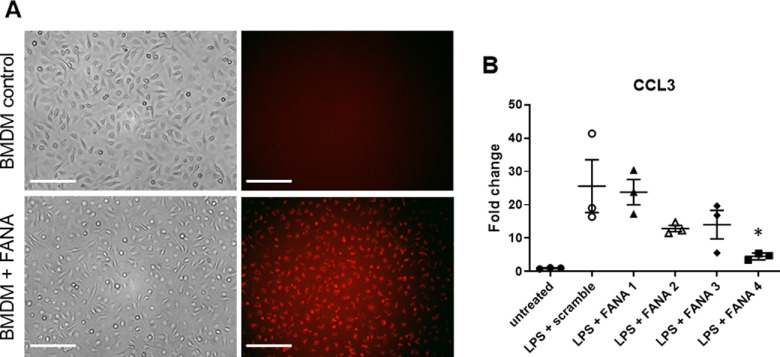
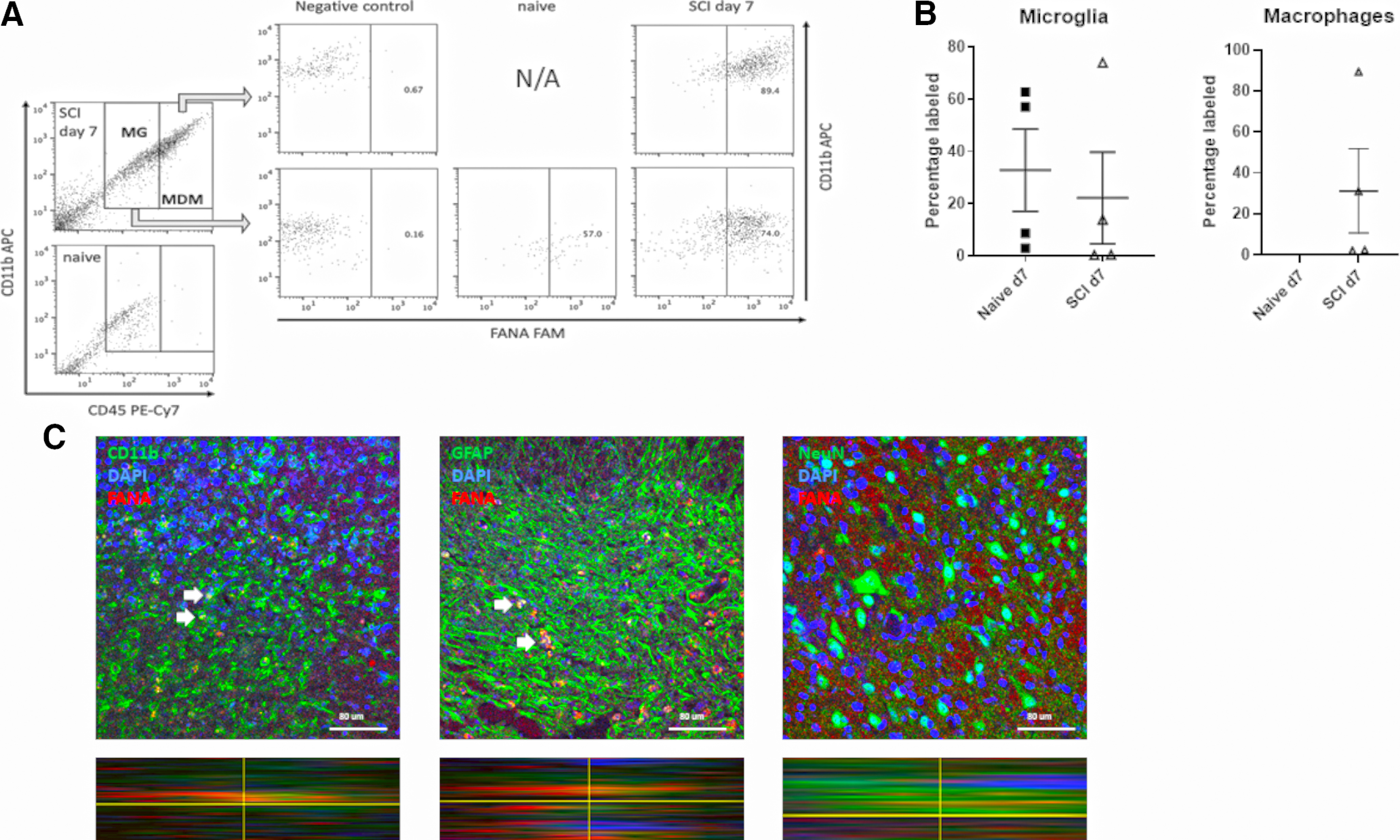
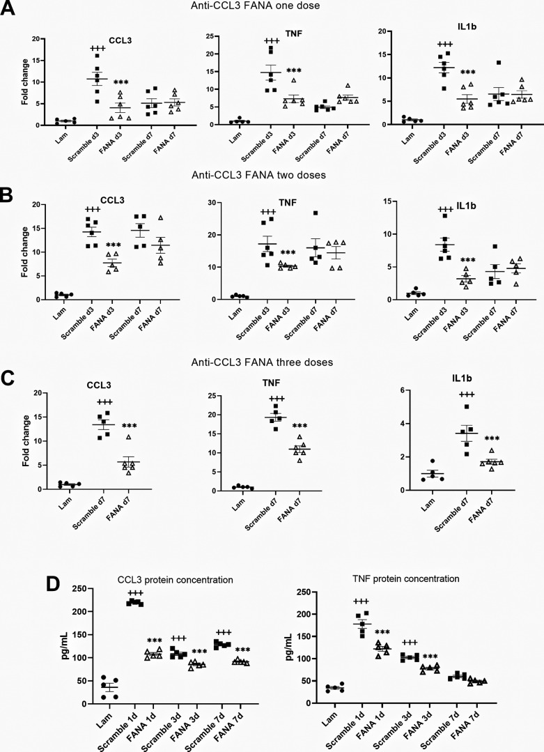
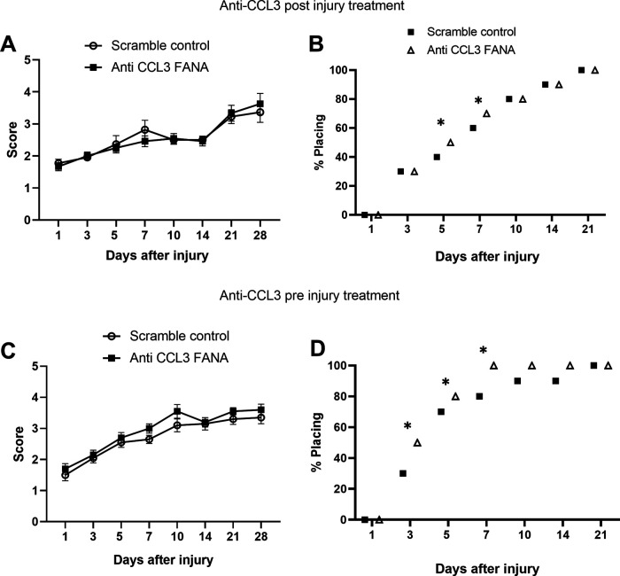
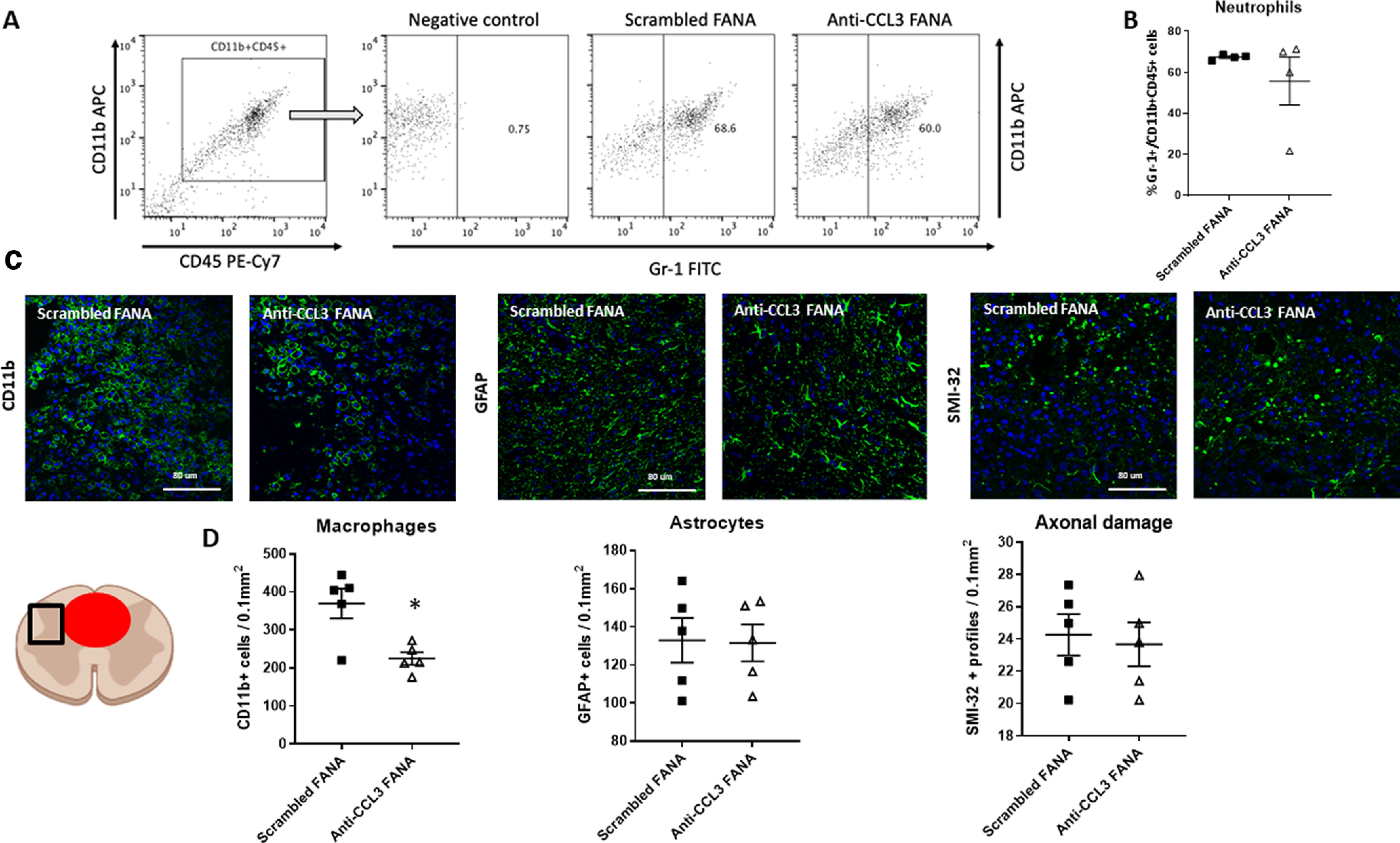
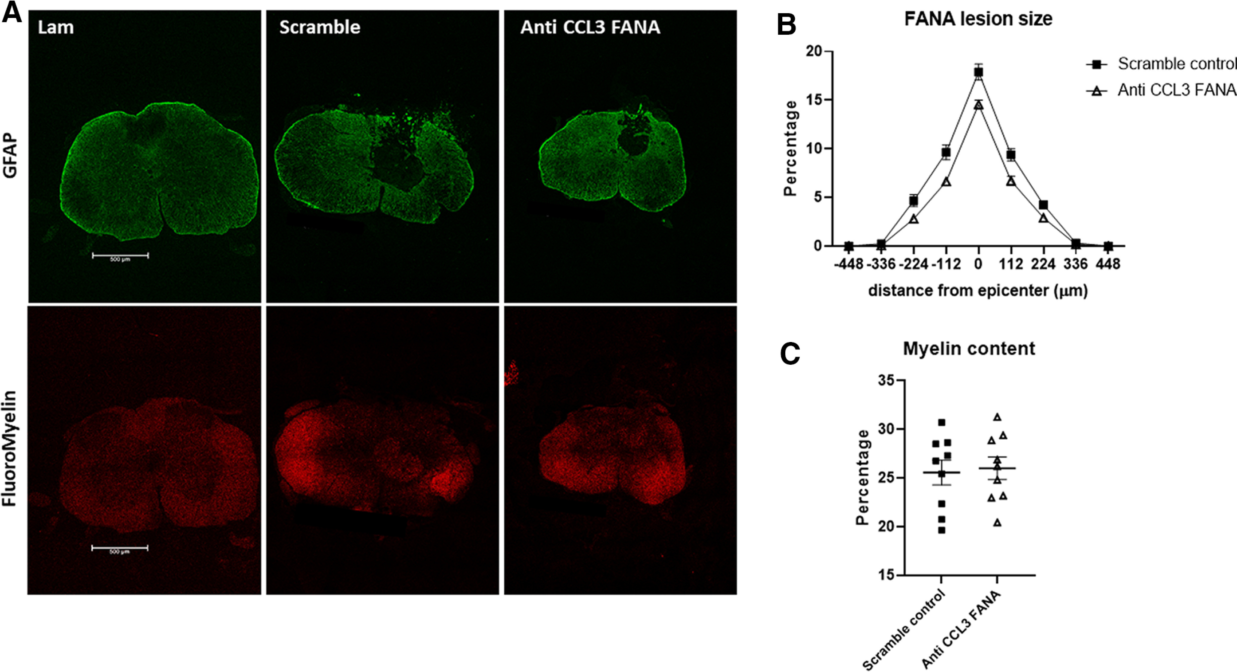
Secondary damage after spinal cord injury (SCI) occurs because of a sequence of events after the initial injury, including exacerbated inflammation that contributes to increased lesion size and poor locomotor recovery. Thus, mitigating secondary damage is critical to preserve neural tissue and improve neurologic outcome. In this work, we examined the therapeutic potential of a novel antisense oligonucleotide (ASO) with special chemical modifications [2'-deoxy-2-fluoro-D-arabinonucleic acid (FANA) ASO] for specifically inhibiting an inflammatory molecule in the injured spinal cord. The chemokine CCL3 plays a complex role in the activation and attraction of immune cells and is upregulated in the injured tissue after SCI. We used specific FANA ASO to inhibit CCL3 in a contusive mouse model of murine SCI. Our results show that self-delivering FANA ASO molecules targeting the chemokine CCL3 penetrate the spinal cord lesion site and suppress the expression of CCL3 transcripts. Furthermore, they reduce other proinflammatory cytokines such as tumor necrosis factor (TNF) and interleukin (IL)-1β after SCI. In summary, we demonstrate for the first time the potential of FANA ASO molecules to penetrate the spinal cord lesion site to specifically inhibit CCL3, reducing proinflammatory cytokines and improve functional recovery after SCI. This novel approach may be used in new treatment strategies for SCI and other pathologic conditions of the CNS.

Keywords: CCL3; FANA ASO; inflammation; novel RNA inhibitor; secondary damage; spinal cord injury.


Figures

Figure 1.
Figure 1.
FANA ASO molecules penetrate BMDMs in a gymnotic fashion and reduce CCL3 expression in vitro. A, When added to the tissue culture media, fluorescently labeled FANA ASO molecules penetrate BMDM cells after 2 h of incubation (red fluorescence, lower panel). The upper panel demonstrates the absence of fluorescence when no FANA ASO molecules were added to the media. Scale bar: 50 μm. B, Assessment of CCL3 knock-down in LPS stimulated BMDMs using four different FANA ASOs molecules. CCL3 mRNA expression levels were compared between LPS-treated BMDMs in the presence of scrambled control FANA ASO or the individual anti-CCL3 FANA ASOs. CCL3 expression levels were unchanged in the presence of FANA-CCL3#1, FANA-CCL3#2, and FANA-CCL3#3. Treatment with FANA-CCL3#4 resulted in a significant reduction of CCL3 expression by ∼80%. Untreated BMDMs are included for reference; n = 3/group; *p < 0.05. Error bars are represented as SEM.
Figure 2.
Figure 2.
Fluorescently labeled FANA ASOs are present at the SCI lesion site after intrathecal injection. A, Representative images of flow cytometry on cells from a section of spinal cord tissue centering on the lesion or spinal cord tissue from naive mice. Green fluorescence derived from FAM-labeled FANA was detectable in gated cell populations for CD11b+/CD45low microglia (MG) and CD11b+/CD45high MDMs. MDM are not detected in naive tissue. B, Quantification of percentages of MG and MDM containing FAM-labeled FANA; n = 4/group, error bars are represented as SEM. C, Seven days after SCI and intrathecal injection, labeled FANA ASO was detectable at the lesion site (red staining). Confocal images with orthogonal view demonstrate uptake of FANA molecules in a non- cell specific manner, as shown by the presence in CD11b+ macrophages/microglia and GFAP+ astrocytes. No uptake was detected in NeuN+ neurons; n = 4–5/group. Scale bars: 80 μm.
Figure 3.
Figure 3.
CCL3 expression levels following SCI were reduced after FANA ASO injection. A, A single injection of FANA ASO CCL3#4 resulted in significant suppression of CCL3 mRNA expression in the injured spinal cord compared with scrambled control at day 3 postinjury. This reduction was accompanied by the suppression of TNF and IL-1β expression, indicating a reduction of the proinflammatory tissue reaction. B, When using a two-injection paradigm, CCL3, TNF, and IL-1β mRNA were also suppressed at day 3. C, When three doses were administered, a sustained suppression of CCL3 was detected at day 7, reducing CCL3 levels by ∼60%. Both TNF and IL-1β gene expression levels were reduced by ∼50%. D, Suppression of CCL3 and TNF in the injured spinal cord was also measured the protein level using ELISA at days 1, 3, and 7 and days 1 and 3 after SCI, respectively; n = 5/group; +++p < 0.001 (compared with laminectomy control); *p < 0.05, ***p < 0.001 (compared with scrambled control of the same time point). Error bars are represented as SEM.
Figure 4.
Figure 4.
Treatment with anti-CCL3 FANA ASO resulted in mild functional improvement. A, BMS scores did not show significant improvement compared with control animals when anti CCL3-FANA was administered three times starting right after injury. B, However, a significantly higher percentage of CCL3-FANA ASO-treated mice performed plantar placement on days 5–7 after injury, compared with control animals. To ensure sufficient time to knock-down CCL3, we also investigated the efficacy of CCL3-FANA ASO pretreatment, with the first dose given 24 h before injury, followed by two more injection immediately after injury and 24 h later. C, BMS scores were not significantly different in treated versus control animals when anti CCL3-FANA was administered three times starting 24 h before injury. D, The pretreatment group, however, showed a significant increase of plantar placement at days 3 through 7 after injury compared with controls; n = 10/group in preinjury treatment paradigm, n = 12/group in postinjury treatment. Data points are represented as mean ± SEM. Note that bars in B, D represent the total percentage of mice from each group capable of plantar placement, thereby not allowing for error bars; *p < 0.05, χ2 test.
Figure 5.
Figure 5.
Treatment with anti-CCL3 FANA ASOs reduced CD11b+ macrophages at the lesion site. A, B. Flow cytometry 1 d after SCI in anti-CCL3-FANA ASO and control-treated animals did not reveal a significant difference in Gr-1+ neutrophils. Numbers were assessed in the CD11b+CD45+ gated cell population. The negative control was stained with CD11b+CD45+ in absence of the Gr-1 antibody. A, Representative flow cytometry images. B, Quantification of CD11b+CD45+Gr-1+ neutrophils. C, Representative images of CD11b+ macrophages/microglia, GFAP+ astrocytes, and SMI-32+ profiles as indicators of axonal damage adjacent to the lesion epicenter in anti-CCL3-FANA ASO and control-treated animals. The schematic indicates the location of the quantified region (square) next to the lesion (red circle). D, Quantification revealed a significant reduction of CD11b+ macrophages/microglia in the anti-CCL3-FANA ASO treatment group, while neither astrocytes nor indicators of axonal damage were different between groups; n = 5/group, data points are represented as mean ± SEM; *p = 0.01. Scale bars: 80 μm.
Figure 6.
Figure 6.
CCL3 inhibition with FANA ASOs did not significantly reduce tissue damage after SCI. Quantification of areas devoid of GFAP immunoreactivity at day 28 after SCI did not show a significant difference in lesion area in anti-CCL3-FANA ASO-pretreated mice compared with control mice. A, Representative images of GFAP and FluoroMyelin stained spinal cord cross sections. B, Quantitative analysis of the lesion volume in CCL3-FANA ASO-pretreated mice and control mice. C, No differences in myelin content, as shown with FluoroMyelin Red staining, were detected between groups; n = 9/group. Data points are represented as mean ± SEM. Scale bars: 500 μm.

References

    1. Baba T, Mukaida N (2014) Role of macrophage inflammatory protein (MIP)-1α/CCL3 in leukemogenesis. Mol Cell Oncol 1:e29899. 10.4161/mco.29899 - DOI - PMC - PubMed
    1. Baby N, Alagappan N, Dheen ST, Sajikumar S (2020) MicroRNA-134-5p inhibition rescues long-term plasticity and synaptic tagging/capture in an Aβ(1-42)-induced model of Alzheimer’s disease. Aging Cell 19:e13046. 10.1111/acel.13046 - DOI - PMC - PubMed
    1. Banisor I, Leist TP, Kalman B (2005) Involvement of beta-chemokines in the development of inflammatory demyelination. J Neuroinflammation 2:7. 10.1186/1742-2094-2-7 - DOI - PMC - PubMed
    1. Bartholdi D, Schwab M (1997) Expression of pro‐inflammatory cytokine and chemokine mRNA upon experimental spinal cord injury in mouse: an in situ hybridization study. Eur J Neurosci 9:1422–1438. 10.1111/j.1460-9568.1997.tb01497.x - DOI - PubMed
    1. Basso DM, Fisher LC, Anderson AJ, Jakeman LB, McTigue DM, Popovich PG (2006) Basso Mouse Scale for locomotion detects differences in recovery after spinal cord injury in five common mouse strains. J Neurotrauma 23:635–659. 10.1089/neu.2006.23.635 - DOI - PubMed

Publication types

Substances

LinkOut - more resources