. 2021 Feb 26;371(6532):910-916.
doi: 10.1126/science.abe0526.

Shulin packages axonemal outer dynein arms for ciliary targeting

Affiliations

Shulin packages axonemal outer dynein arms for ciliary targeting

Girish R Mali et al. Science. .

Abstract

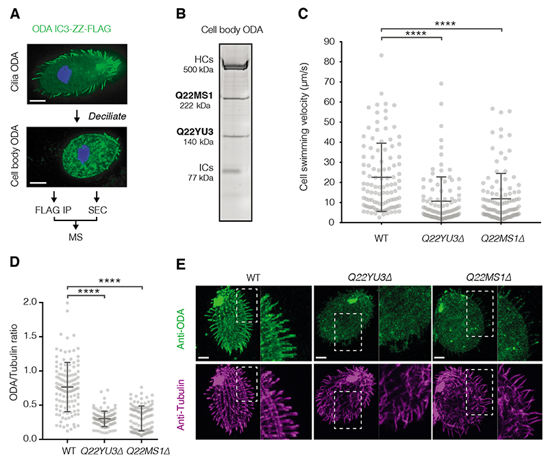
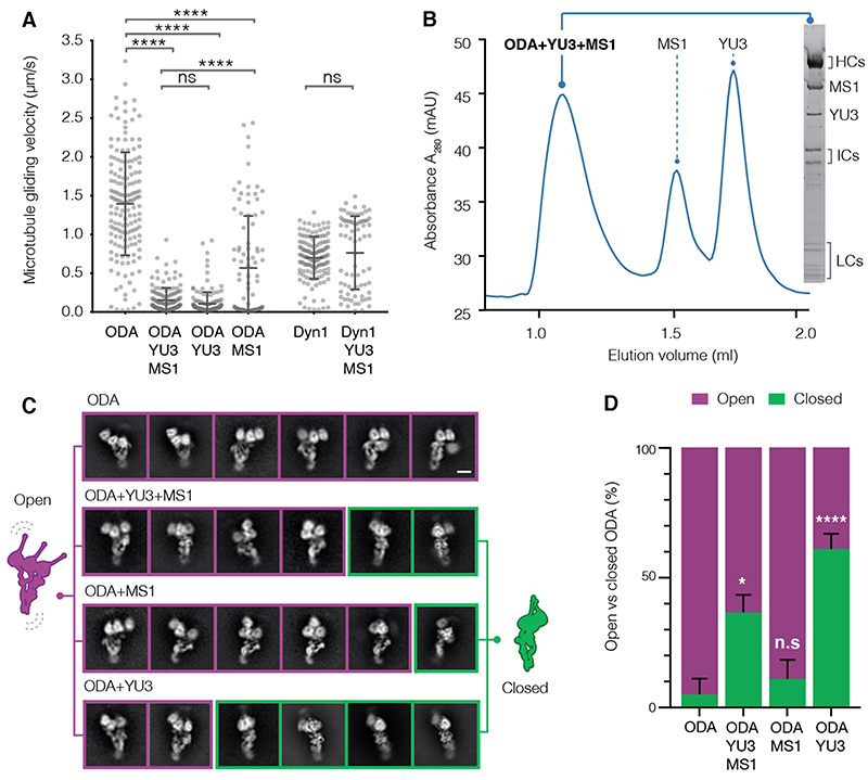
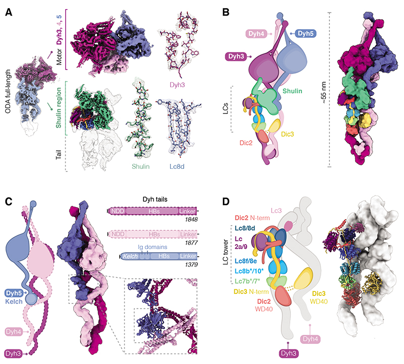
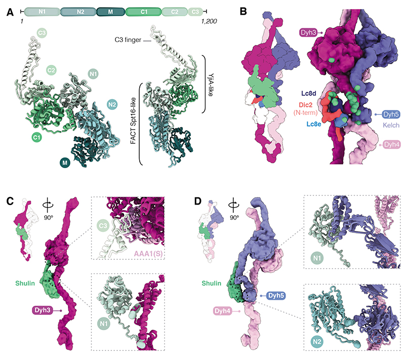
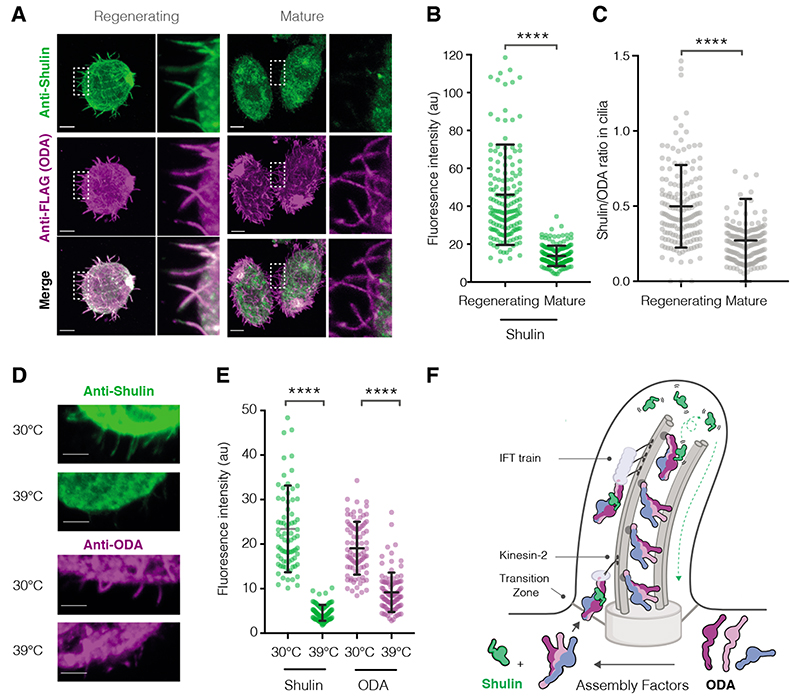
The main force generators in eukaryotic cilia and flagella are axonemal outer dynein arms (ODAs). During ciliogenesis, these ~1.8-megadalton complexes are assembled in the cytoplasm and targeted to cilia by an unknown mechanism. Here, we used the ciliate Tetrahymena to identify two factors (Q22YU3 and Q22MS1) that bind ODAs in the cytoplasm and are required for ODA delivery to cilia. Q22YU3, which we named Shulin, locked the ODA motor domains into a closed conformation and inhibited motor activity. Cryo-electron microscopy revealed how Shulin stabilized this compact form of ODAs by binding to the dynein tails. Our findings provide a molecular explanation for how newly assembled dyneins are packaged for delivery to the cilia.


Conflict of interest statement

Competing interests The authors declare no competing interests.

Figures

Fig. 1
Fig. 1. Q22MS1 and Q22YU3 deliver ODAs from cytoplasm to cilia
(A) Scheme used to identify novel interactors of ODAs assembled in the cell body. (B) SDS-PAGE of ODA purified from the cell body by IP-SEC showing co-elution with Q22MS1 and Q22YU3, HC: Heavy chains, IC: Intermediate chains. (C) Cell swimming velocity comparing wildtype (WT n=108) and mutant strains (Q22YU3Δ n=102 and Q22MS1Δ n=110). (D) Ratio of ODA/Tubulin immunofluorescence intensity along individual cilia (WT n=118, Q22YU3Δ n=104, Q22MS1Δ n=129, 3-10 cilia from 14-17 cells/genotype). (E) Representative cells showing immunofluorescence for ODA and tubulin (quantified in D). Insets highlight reduced amount of ODA staining in mutant cilia compared to wildtype cilia. Scale bars:10 μm. Error bars show standard deviation; ns=not significantly different, ****p≤ 0.0001 (ANOVA with Tukey’s test for multiple comparisons).
Fig. 2
Fig. 2. Q22YU3 binding inhibits ODAs by clustering motors
(A) Microtubule gliding velocities. Individual gliding events from three technical replicates/sample are plotted (ODA n=159, ODA+YU3+MS1 n=136, ODA+YU3 n=146, ODA+MS1 n=94, Dyn1 n=136, Dyn1+YU3+MS1 n=76). YU3: Q22YU3, MS1: Q22MS1, Dyn1: human cytoplasmic dynein 1. Error bars show standard deviation; ns=not significantly different, ****p≤ 0.0001 (ANOVA with Tukey’s test for multiple comparisons). (B) Axonemal-purified open ODA reconstituted with recombinant Q22YU3 and Q22MS1. SDS-PAGE gel of SEC peak fraction. (C) Representative 2D class averages showing the distribution of open (purple) and closed (green) ODA particles from ODAs alone and reconstituted with factors. Scale bars = 10 nm. (D) Quantification of closed and open particles from 18,000 particles/dataset, (n=3 technical replicates) represented as percentage. Error bars show standard deviation. ns=not significantly different, * p≤ 0.01, ****p≤ 0.0001 (Kruskal Wallis test). Refer to Fig. S6 and Table S5.
Fig. 3
Fig. 3. Cryo-EM reconstruction shows architecture of closed ODA
(A) Overview of the closed ODA bound by Shulin with head (purple) and tail (blue) maps docked in an overall map (grey). Maps obtained after masked refinements are shown for the head region containing densities for Dyh3, 4 and 5 motor domains and the ordered tail map contains a docked Shulin-region map (green). Representative cryo-EM densities are shown. The following contour levels (σ) are used; right panel-ODA full-length structure: 0.000607, tail: 0.0026, motors: 0.01; middle panel-ordered tail: 0.00448, Shulin region: 0.015, motors: Dyh3 0.01, Dyh4 0.015, Dyh5 0.015. Zoom insets showing side chain densities were at contour level of 0.02. (B) Cartoon and filtered surface representation of all modeled subunits. (C) Dyh5 binds Dyh4 via its N-terminal Kelch domain (inset). HB: Helical bundles, NDD: N-terminal dimerization domain. (D) DIC N-terminal extensions bind dimers of LCs forming a LC tower and followed by globular WD40 domains that contact Dyh3 and Dyh4. Heterodimers of Lc7b/7 and Lc8b/Lc10 are tentatively assigned (*). Lc3 sits on Dyh4 and is not part of the LC tower.
Fig. 4
Fig. 4. Characterization of Shulin structure and its mechanism of ODA inhibition
(A) Domain architecture of Shulin. N-terminal (N1, N2) and Middle (M) domains share a similar fold with the FACT complex core subunit Spt16. C-terminal (C1, C2) domains are similar to GTPase YjiA and are followed by a C-terminal finger (C3). (B) Cartoon and filtered surface representation with main contacts between Shulin and ODA subunits highlighted in green spheres. (C) Shulin’s N1 domain contacts helical bundles proximal to the linker in Dyh3 tail. The C3 finger projects out to contact Dyh3 AAA1(S) (insets). (D) Shulin’s N1 domain contacts Dyh5 helical bundles and its N2 domain touches the Kelch domain. Shulin contacts Dyh4 just below Dyh5 Kelch-domain (insets).
Fig. 5
Fig. 5. Shulin delivers ODAs to regenerating cilia
(A) Co-immunostaining of ODAs (anti-FLAG) and Shulin in the IC3-FLAG strain. Representative cells regenerating cilia 30-minutes post-deciliation and with mature cilia are shown, insets highlight differences in levels of Shulin and constant levels of ODA in cilia in both conditions. (B, C) Quantification of Shulin levels in regenerating and mature cilia and ratios of per cilium fluorescence intensities for Shulin/ODA (Regenerating n=160, Mature n=162, 9-10 cilia from 16 cells/condition). (D, E) Immunostaining for Shulin and ODA (anti-ODA) in an ODA temperature sensitive mutant strain (OAD1C11) grown at permissive (30°C) and restrictive (39°C) temperatures for 16 hours (Shulin: 30°C n=78, 39°C n=83; ODA: 30°C n=87, 39°C n=95 cilia from 10 cells per condition). Scale bars:10 μm. Error bars show standard deviation, ****p≤ 0.0001 (ANOVA). (F) Model for Shulin’s role in delivering ODAs to cilia. Shulin locks ODAs in their closed state during IFT (intraflagellar transport) in regenerating cilia. Shulin is released after ODAs stably dock and leaves the ciliary compartment (green arrow)

References

    1. Drummond IA. Cilia functions in development. Curr Opin Cell Biol. 2012;24:24–30. - PMC - PubMed
    1. Goodenough UW, Heuser JE. Outer and inner dynein arms of cilia and flagella. Cell. 1985;41:341–342. - PubMed
    1. Leigh MW, Pittman JE, Carson JL, Ferkol TW, Dell SD, Davis SD, Knowles MR, Zariwala Ma. Clinical and genetic aspects of primary ciliary dyskinesia/Kartagener syndrome. Genet Med. 2009;11:473–487. - PMC - PubMed
    1. King SM. Axonemal dynein arms. Cold Spring Harb Perspect Biol. 2016;8:a028100. - PMC - PubMed
    1. Fowkes ME, Mitchell DR. The Role of Preassembled Cytoplasmic Complexes in Assembly of Flagellar Dynein Subunits. Mol Biol Cell. 1998;9:2337–2347. - PMC - PubMed

Publication types

MeSH terms