Clinical Trial
. 2021 Feb 25;12(1):1178.
doi: 10.1038/s41467-021-21371-5.

Lentivirus-mediated gene therapy for Fabry disease

Affiliations
Clinical Trial

Lentivirus-mediated gene therapy for Fabry disease

Aneal Khan et al. Nat Commun. .

Abstract

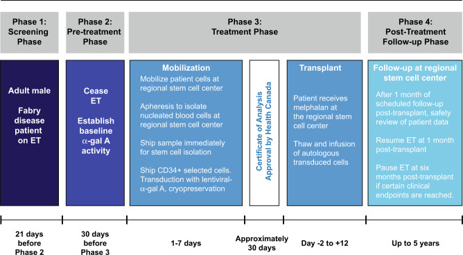
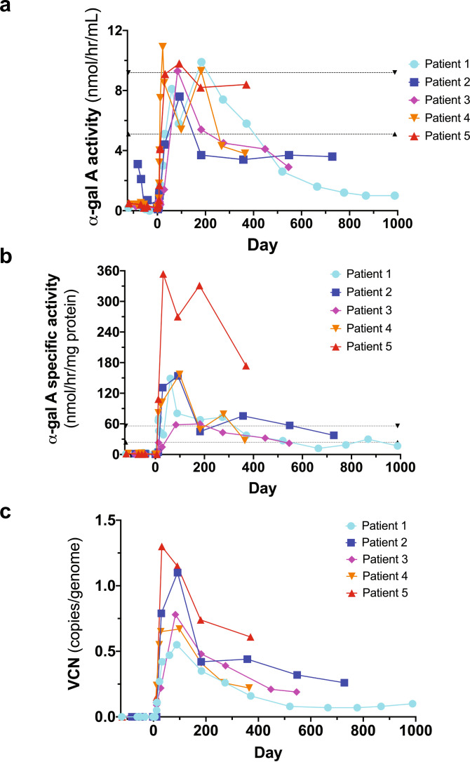
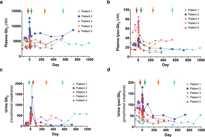
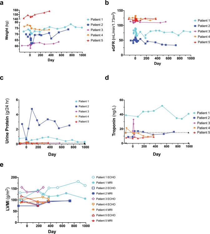
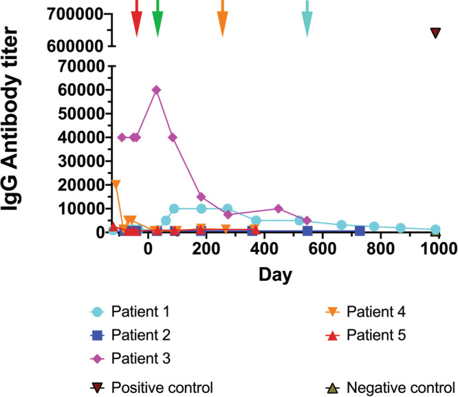
Enzyme and chaperone therapies are used to treat Fabry disease. Such treatments are expensive and require intrusive biweekly infusions; they are also not particularly efficacious. In this pilot, single-arm study (NCT02800070), five adult males with Type 1 (classical) phenotype Fabry disease were infused with autologous lentivirus-transduced, CD34+-selected, hematopoietic stem/progenitor cells engineered to express alpha-galactosidase A (α-gal A). Safety and toxicity are the primary endpoints. The non-myeloablative preparative regimen consisted of intravenous melphalan. No serious adverse events (AEs) are attributable to the investigational product. All patients produced α-gal A to near normal levels within one week. Vector is detected in peripheral blood and bone marrow cells, plasma and leukocytes demonstrate α-gal A activity within or above the reference range, and reductions in plasma and urine globotriaosylceramide (Gb3) and globotriaosylsphingosine (lyso-Gb3) are seen. While the study and evaluations are still ongoing, the first patient is nearly three years post-infusion. Three patients have elected to discontinue enzyme therapy.


Conflict of interest statement

A.K. received grants, consulting fees, revenue distribution agreement, speaker fees and travel support with AVROBIO, Inc. as well as revenue distribution agreement with University Health Network regarding gene therapy using technology from this work. D.L.B. and J.H. were partially paid from a Sponsored Research Agreement—AVROBIO, Inc. C. A. Rupar has the following financial relationships to disclose: the Biochemical Genetics clinical diagnostic laboratory at his home institution is contracted by AVROBIO, Inc. to assay enzymes on a fee for service basis. He is the laboratory director but receives no personal compensation. C.A.-B. has received a service contract and honoraria for biomarker analysis with AVROBIO, Inc., grant from CIHR. K.G. had travel paid for by AVROBIO, Inc. S.W. has received nonfinancial support from Sanofi-Genzyme, nonfinancial support from Takeda Pharmaceuticals (formerly Shire HGT), personal fees and nonfinancial support from Amicus Therapeutics. K.L. has received travel grant and honorarium from Amicus Therapies; travel grant and speaker fees from Sanofi-Genzyme; travel grant, consulting fees and speaker fees from Takeda Pharmaceuticals; medical advisor to the Canadian Fabry Disease Association. C.F.M. has received grants, personal fees, and nonfinancial support from Takeda Pharmaceuticals (previously Shire HGT), grants, personal fees and nonfinancial support from Sanofi-Genzyme, nonfinancial support from Amicus Therapeutics. A.K. has received consultancy fees from AVROBIO, Inc. unrelated to this study. M.L.W. has received research grants, consulting fees, speaker fees and travel support with Amicus Therapeutics, Protalix, Sanofi-Genzyme and Takeda, revenue distribution agreement with University Health Network regarding gene therapy using technology from this work. J.A.M. has the following financial relationships to disclose: SAB—Rapa Therapeutics. Honoraria—Sanofi-Genzyme, Shire. Co-Founder—AVROBIO, Inc. Shareholder—AVROBIO, Inc. Grants from Canadian Institutes of Health Research and Kidney Foundation of Canada and AVROBIO, Inc. M.B., A.C., P.R.D., R.F., D.H.F., G.F., S.J., W.M.M., P.O., N.P., and J.W.R. have no financial relationships to disclose in relation to this trial.

Figures

Fig. 1
Fig. 1. Study schema.
Five men with Type I Fabry disease were infused with autologous transduced CD34+-selected hematopoietic stem/progenitor cells engineered to express α-galactosidase A (α-gal A) following mild ablation. ET enzyme therapy.
Fig. 2
Fig. 2. α-galactosidase A (α-gal A) enzyme activity and vector copy number (VCN).
a Plasma α-gal A activity attained reference range levels in all patients; although decreased over time, the plasma α-gal A enzyme activity levels are above what is observed in Fabry disease patients and have not returned to original baseline levels. The reference ranges (dotted lines) were defined by Dr. Rupar’s laboratory based on 150 specimens referred for diagnostic testing. Males with classic Fabry disease have plasma levels around 1 nmol/h/ml. b Leukocyte α-gal A attained supranormal specific activity levels for each patient. Although decreased over time, leukocyte α-gal A-specific enzyme activity levels are above what is seen in Fabry disease patients and have not returned to original baseline levels. The reference ranges (dotted lines) were defined by Dr. Rupar’s laboratory based on 150 specimens referred for diagnostic testing. c VCN in peripheral blood reached between 0.55 and 1.10 copies/genome in all patients, and although decreased over time, has remained above 0.05 copies/genome in all patients to date (almost 3 years in Patient 1).
Fig. 3
Fig. 3. Total plasma and urine globotriaosylceramide (Gb3) and lyso-Gb3 levels.
Plasma Gb3 (a), plasma lyso-Gb3 (b), urine Gb3 (c), and urine lyso-Gb3 (d) levels are illustrated for each patient. Red arrow is at Day −30 when ET was stopped prior to mobilization. Green arrow is at Day 30 when ET was restarted for Patients 1, 2, 4, and 5. The orange arrow demarks when Patient 4 stopped ET at Day 214 and the blue arrow is when Patient 1 stopped ET at Day 548. Patient 3 chose not to restart ET.
Fig. 4
Fig. 4. Clinical parameters.
Weight (a), estimated Glomerular Filtration Rate (eGFR) (b), urinary protein secretion (c), troponin (d), and Left Ventricular Mass Index (LVMI) (e) was monitored during the course of the trial for all patients. eGFR was calculated using the Chronic Kidney Disease-Epidemiology Collaboration formula. Values from Magnetic Resonance Imaging (MRI) and Echocardiography (ECHO) are shown for LVMI (e). Patient 3 did not attend all cardiac assessments and has been omitted from Fig. 4e.
Fig. 5
Fig. 5. Immunoglobulin G (IgG) antibody titer.
Enzyme-Linked Immunosorbent Assays (ELISA) were completed at each time point as shown. In four patients who had detectable anti- α-galactosidase A (α-gal A) antibodies, levels declined in three patients and rose in one patient, although the titers never reached the levels of a positive control sample collected following enzyme therapy (ET). Red arrow is at Day −30 when ET was stopped prior to mobilization. Green arrow is at Day 30 when ET was restarted for Patients 1, 2, 4, and 5. The orange arrow demarks when Patient 4 stopped ET at Day 214 and the blue arrow is when Patient 1 stopped ET at Day 548. Patient 3 chose not to restart ET.

References

    1. Brady RO, et al. Enzymatic defect in Fabry’s disease. Ceramidetrihexosidase deficiency. N. Engl. J. Med. 1967;276:1163–1167. doi: 10.1056/NEJM196705252762101. - DOI - PubMed
    1. Platt FM, d’Azzo A, Davidson BL, Neufeld EF, Tifft CJ. Lysosomal storage diseases. Nat. Rev. Dis. Prim. 2018;4:27. doi: 10.1038/s41572-018-0025-4. - DOI - PubMed
    1. Aerts JM, et al. Elevated globotriaosylsphingosine is a hallmark of Fabry disease. Proc. Natl Acad. Sci. USA. 2008;105:2812–2817. doi: 10.1073/pnas.0712309105. - DOI - PMC - PubMed
    1. Auray-Blais C, et al. Urinary globotriaosylsphingosine-related biomarkers for Fabry disease targeted by metabolomics. Anal. Chem. 2012;84:2745–2753. doi: 10.1021/ac203433e. - DOI - PubMed
    1. Desnick RJ, et al. Fabry disease, an under-recognized multisystemic disorder: expert recommendations for diagnosis, management, and enzyme replacement therapy. Ann. Intern. Med. 2003;138:338–346. doi: 10.7326/0003-4819-138-4-200302180-00014. - DOI - PubMed

Publication types

Associated data

Grants and funding