. 2021 Feb 25;11(1):4666.
doi: 10.1038/s41598-021-83849-y.

A matched irrigation and obturation strategy for root canal therapy

Affiliations

A matched irrigation and obturation strategy for root canal therapy

Rafaela Fernandes Zancan et al. Sci Rep. .

Abstract

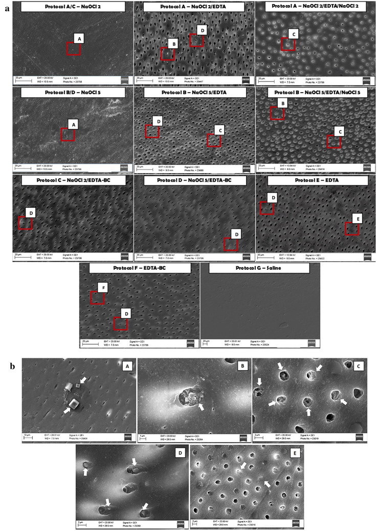
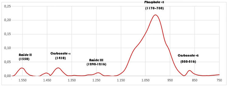
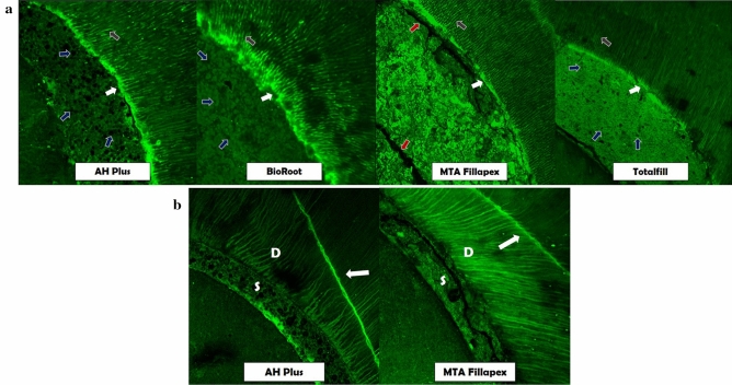
In root canal therapy, irrigating solutions are employed to eliminate the bacterial load and also prepare dentin for sealer interaction. The aim of this research was to assess how irrigating solutions employed on their own or in sequence affected the tooth structure. The best way to prepare the tooth for obturation using hydraulic calcium silicate cement (HCSC) sealers and gutta-percha, thus guiding clinicians on a matched irrigation-obturation strategy for optimized root canal treatment was investigated. The effect of irrigating solutions on dentine was investigated by assessing changes in dentin microhardness, ultrastructure and mineral content, organic/inorganic matter, surface roughness and Young's modulus. The interaction of four root canal sealers with the dentin was analysed by assessing the changes in microhardness of the dentin after sealer placement and also the sealer to dentin interface by scanning electron and confocal laser microscopy. The irrigating solutions damaged the dentin irreversibly both when used on their own and in combination. The best sequence involved sodium hypochlorite followed by chelator and a final rinse with sodium hypochlorite and obturation using HCSC sealers that enabled the restoration of dentin properties. The HCSC sealers did not rely on chelator irrigating solutions for a good material adaptation to dentin.


Conflict of interest statement

The authors declare no competing interests.

Figures

Figure 1
Figure 1
(a) Representative images of root dentin at × 2 K magnification after the different irrigation protocols on the surface. The letters on the images indicate the areas that were viewed at higher magnification and are shown in (b). (b) High magnification scanning electron micrographs (3 K ×: A,C,E; 4 K ×: B,D); of the areas marked by the letters A–E in Figure (a). The areas are indicated with white arrows. (A,B) shows the presence of cuboid crystals rich in sodium and chlorine on dentin and inside dentinal tubules, respectively; (C) shows the peritubular and intertubular dentinal erosion; (D) shows the presence of smear layer inside dentinal tubules and (E) shows the sclerosis of dentinal tubules.
Figure 2
Figure 2
Representative infrared spectral region between 1600 and 750 cm−1 of untreated dentin showing the absorption peaks of the main dentin components.
Figure 3
Figure 3
Atomic force micrographs showing topography of the dentin surface before (initial—I) and after immersion in the irrigation solutions (G1–G11; horizontal axis) as indicated in Protocols A–G detailed in Table 1. White arrows indicate less evident dentinal tubules in groups with NaOCl or chelator single flush. Light grey and dark grey arrows expressed a wider range and progressive widening of the dentinal tubules when both irrigants were used in a Protocol alone or adding NaOCl as a final flush, respectively. In protocol NaOCl+ chelator, (5%) NaOCl promoted a rough surface of dentin indicated by red arrows.
Figure 4
Figure 4
Representative fluorescence-mode CLSM images of sealer-dentin interface demonstrated by Orange G added to the test sealers; (a) showing a richly dye-infiltrated layer at the sealer-dentin interface marked with white arrows, sealer penetration inside dentinal tubules marked with grey arrows, porosity of sealers with blue arrows and cracks on sealer mass with red arrows; (b) cracks indicated by fluorescent radial lines on dentin (D) marked by white arrows and on the sealer (S) for MTA Fillapex indicated with red arrows.

Similar articles

Cited by

References

    1. Ma J, Wang Z, Shen Y, Haapasalo M. A new noninvasive model to study the effectiveness of dentin disinfection by using confocal laser scanning microscopy. J. Endod. 2011;37:1380–1385. doi: 10.1016/j.joen.2011.06.018. - DOI - PubMed
    1. Wang Z, Shen Y, Haapasalo M. Effectiveness of endodontic disinfecting solutions against young and old Enterococcus faecalis biofilms in dentin canals. J. Endod. 2012;38:1376–1379. doi: 10.1016/j.joen.2012.06.035. - DOI - PubMed
    1. Huang Y, et al. Micro-CT and nano-CT analysis of filling quality of three different endodontic sealers. Dentomaxillofac. Radiol. 2017;46:20170223. doi: 10.1259/dmfr.20170223. - DOI - PMC - PubMed
    1. Çalt S, Serper A. Time-dependent effects of EDTA on dentin structures. J. Endod. 2002;28:17–19. doi: 10.1097/00004770-200201000-00004. - DOI - PubMed
    1. Haapasalo M, Shen Y, Qian W, Gao Y. Irrigation in endodontics. Dent. Clin. N. Am. 2010;54:291–312. doi: 10.1016/j.cden.2009.12.001. - DOI - PubMed

Publication types

LinkOut - more resources