

Solidified glomerulosclerosis, identified using single glomerular proteomics, predicts end-stage renal disease in Chinese patients with type 2 diabetes
- PMID: 33633132
- PMCID: PMC7907371
- DOI: 10.1038/s41598-021-83856-z
Solidified glomerulosclerosis, identified using single glomerular proteomics, predicts end-stage renal disease in Chinese patients with type 2 diabetes
Abstract
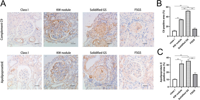
Few histological prognostic indicators for end-stage renal disease (ESRD) have been validated in diabetic patients. This biopsy-based study aimed to identify nephropathological risk factors for ESRD in Chinese patients with type 2 diabetes. Histological features of 322 Chinese type 2 diabetic patients with biopsy-confirmed diabetic nephropathy (DN) were retrospectively analysed. Cox proportional hazards analysis was used to estimate the hazard ratio (HR) for ESRD. Single glomerular proteomics and immunohistochemistry were used to identify differentially expressed proteins and enriched pathways in glomeruli. During the median follow-up period of 24 months, 144 (45%) patients progressed to ESRD. In multivariable models, the Renal Pathology Society classification failed to predict ESRD, although the solidified glomerulosclerosis (score 1: HR 1.65, 95% confidence interval [CI] 1.04-2.60; score 2: HR 2.48, 95% CI 1.40-4.37) and extracapillary hypercellularity (HR 2.68, 95% CI 1.55-4.62) were identified as independent risk factors. Additionally, single glomerular proteomics, combined with immunohistochemistry, revealed that complement C9 and apolipoprotein E were highly expressed in solidified glomerulosclerosis. Therefore, solidified glomerulosclerosis and extracapillary hypercellularity predict diabetic ESRD in Chinese patients. Single glomerular proteomics identified solidified glomerulosclerosis as a unique pathological change that may be associated with complement overactivation and abnormal lipid metabolism.
Conflict of interest statement
The authors declare no competing interests.
Figures


Similar articles
-
Urinary complement proteins and risk of end-stage renal disease: quantitative urinary proteomics in patients with type 2 diabetes and biopsy-proven diabetic nephropathy.J Endocrinol Invest. 2021 Dec;44(12):2709-2723. doi: 10.1007/s40618-021-01596-3. Epub 2021 May 27. J Endocrinol Invest. 2021. PMID: 34043214 Free PMC article.
-
DIABETIC RETINOPATHY, CLASSIFIED USING THE LESION-AWARE DEEP LEARNING SYSTEM, PREDICTS DIABETIC END-STAGE RENAL DISEASE IN CHINESE PATIENTS.Endocr Pract. 2020 Apr;26(4):429-443. doi: 10.4158/EP-2019-0512. Epub 2020 Jan 22. Endocr Pract. 2020. PMID: 31968187
-
Clinicopathologic features and prognostic factors in older patients with biopsy-proven diabetic nephropathy.Int Urol Nephrol. 2021 Jun;53(6):1161-1170. doi: 10.1007/s11255-020-02710-9. Epub 2021 Jan 3. Int Urol Nephrol. 2021. PMID: 33389518
-
Segmental Sclerosis and Extracapillary Hypercellularity Predict Diabetic ESRD.J Am Soc Nephrol. 2018 Feb;29(2):694-703. doi: 10.1681/ASN.2017020192. Epub 2017 Nov 27. J Am Soc Nephrol. 2018. PMID: 29180393 Free PMC article.
-
Lessons learned from studies of the natural history of diabetic nephropathy in young type 1 diabetic patients.Pediatr Endocrinol Rev. 2008 Aug;5 Suppl 4:958-63. Pediatr Endocrinol Rev. 2008. PMID: 18806710 Review.
Cited by
-
Association of the podocyte phenotype with extracapillary hypercellularity in patients with diabetic kidney disease.J Nephrol. 2024 Nov;37(8):2209-2222. doi: 10.1007/s40620-024-01981-0. Epub 2024 Jul 27. J Nephrol. 2024. PMID: 39066994
-
Urinary complement proteins and risk of end-stage renal disease: quantitative urinary proteomics in patients with type 2 diabetes and biopsy-proven diabetic nephropathy.J Endocrinol Invest. 2021 Dec;44(12):2709-2723. doi: 10.1007/s40618-021-01596-3. Epub 2021 May 27. J Endocrinol Invest. 2021. PMID: 34043214 Free PMC article.
-
Metabolic phenotypes and risk of end-stage kidney disease in patients with type 2 diabetes.Front Endocrinol (Lausanne). 2023 May 10;14:1103251. doi: 10.3389/fendo.2023.1103251. eCollection 2023. Front Endocrinol (Lausanne). 2023. PMID: 37234807 Free PMC article.
-
Holistic fine-grained global glomerulosclerosis characterization: from detection to unbalanced classification.J Med Imaging (Bellingham). 2022 Jan;9(1):014005. doi: 10.1117/1.JMI.9.1.014005. Epub 2022 Feb 17. J Med Imaging (Bellingham). 2022. PMID: 35237706 Free PMC article.
-
Insights into the molecular underpinning of type 2 diabetes complications.Hum Mol Genet. 2025 Mar 7;34(6):469-480. doi: 10.1093/hmg/ddae203. Hum Mol Genet. 2025. PMID: 39807636 Free PMC article. Review.
References
Publication types
MeSH terms
LinkOut - more resources
Full Text Sources
Other Literature Sources
Medical
Research Materials
Miscellaneous
