

Airspace Dimension Assessment (AiDA) by inhaled nanoparticles: benchmarking with hyperpolarised 129Xe diffusion-weighted lung MRI
- PMID: 33633165
- PMCID: PMC7907057
- DOI: 10.1038/s41598-021-83975-7
Airspace Dimension Assessment (AiDA) by inhaled nanoparticles: benchmarking with hyperpolarised 129Xe diffusion-weighted lung MRI
Abstract
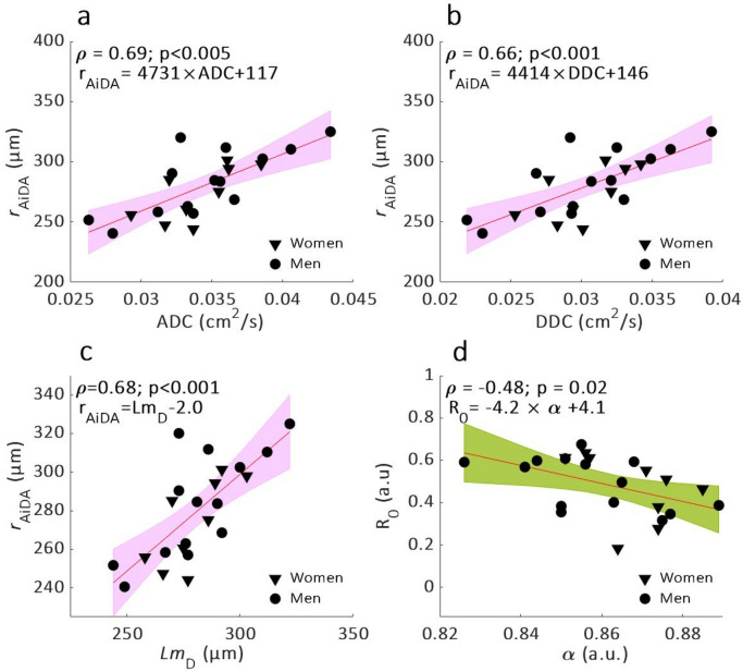
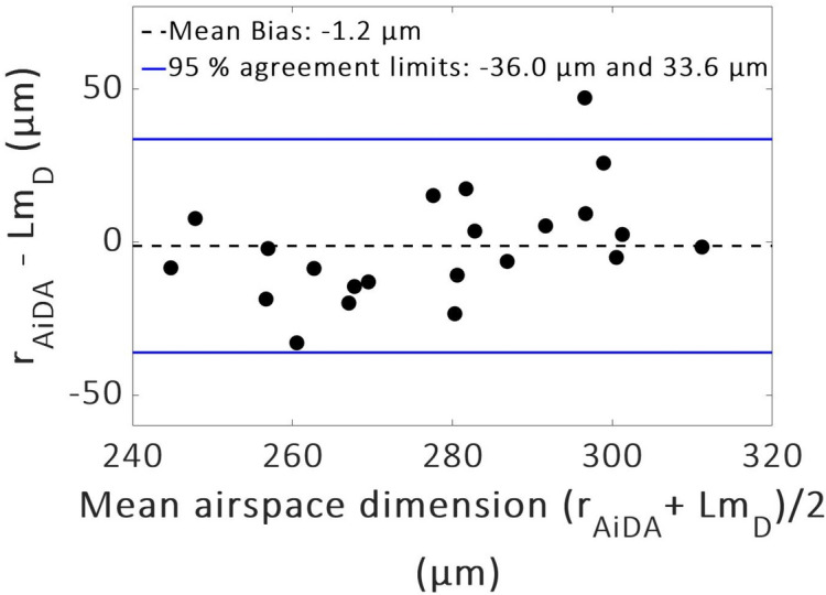
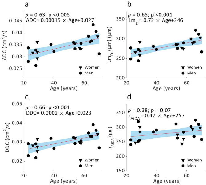
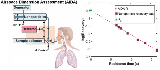
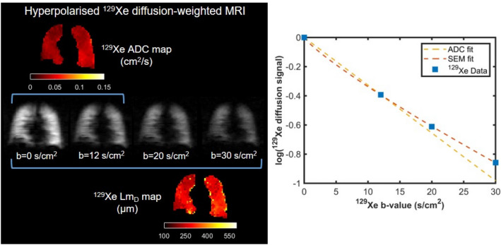
Enlargements of distal airspaces can indicate pathological changes in the lung, but accessible and precise techniques able to measure these regions are lacking. Airspace Dimension Assessment with inhaled nanoparticles (AiDA) is a new method developed for in vivo measurement of distal airspace dimensions. The aim of this study was to benchmark the AiDA method against quantitative measurements of distal airspaces from hyperpolarised 129Xe diffusion-weighted (DW)-lung magnetic resonance imaging (MRI). AiDA and 129Xe DW-MRI measurements were performed in 23 healthy volunteers who spanned an age range of 23-70 years. The relationship between the 129Xe DW-MRI and AiDA metrics was tested using Spearman's rank correlation coefficient. Significant correlations were observed between AiDA distal airspace radius (rAiDA) and mean 129Xe apparent diffusion coefficient (ADC) (p < 0.005), distributed diffusivity coefficient (DDC) (p < 0.001) and distal airspace dimension (LmD) (p < 0.001). A mean bias of - 1.2 µm towards rAiDA was observed between 129Xe LmD and rAiDA, indicating that rAiDA is a measure of distal airspace dimension. The AiDA R0 intercept correlated with MRI 129Xe α (p = 0.02), a marker of distal airspace heterogeneity. This study demonstrates that AiDA has potential to characterize the distal airspace microstructures and may serve as an alternative method for clinical examination of the lungs.
Conflict of interest statement
Prof. P. Wollmer received personal fees from Chiesi Pharmaceuticals during the conduct of the study. In addition, Prof. P Wollmer and Assoc. Prof. J. Löndahl have a patent for “Device and Method for pulmonary function measurements” issued. The other authors declare no competing interests.
Figures
Similar articles
-
Airspace Dimension Assessment with Nanoparticles (AiDA) in Comparison to Established Pulmonary Function Tests.Int J Nanomedicine. 2022 Jun 25;17:2777-2790. doi: 10.2147/IJN.S360271. eCollection 2022. Int J Nanomedicine. 2022. PMID: 35782019 Free PMC article.
-
Charting the human respiratory tract with airborne nanoparticles: evaluation of the Airspace Dimension Assessment technique.J Appl Physiol (1985). 2018 Dec 1;125(6):1832-1840. doi: 10.1152/japplphysiol.00410.2018. Epub 2018 Sep 6. J Appl Physiol (1985). 2018. PMID: 30188799
-
Airspace Dimension Assessment with nanoparticles reflects lung density as quantified by MRI.Int J Nanomedicine. 2018 May 21;13:2989-2995. doi: 10.2147/IJN.S160331. eCollection 2018. Int J Nanomedicine. 2018. PMID: 29861632 Free PMC article.
-
Diffusion lung imaging with hyperpolarized gas MRI.NMR Biomed. 2017 Mar;30(3):10.1002/nbm.3448. doi: 10.1002/nbm.3448. Epub 2015 Dec 16. NMR Biomed. 2017. PMID: 26676342 Free PMC article. Review.
-
Hyperpolarized gas diffusion MRI for the study of atelectasis and acute respiratory distress syndrome.NMR Biomed. 2014 Dec;27(12):1468-78. doi: 10.1002/nbm.3136. Epub 2014 Jun 11. NMR Biomed. 2014. PMID: 24920074 Free PMC article. Review.
Cited by
-
Airspace Dimension Assessment with Nanoparticles (AiDA) in Comparison to Established Pulmonary Function Tests.Int J Nanomedicine. 2022 Jun 25;17:2777-2790. doi: 10.2147/IJN.S360271. eCollection 2022. Int J Nanomedicine. 2022. PMID: 35782019 Free PMC article.
-
Diffusion weighted hyperpolarized 129 Xe MRI of the lung with 2D and 3D (FLORET) spiral.Magn Reson Med. 2023 Apr;89(4):1342-1356. doi: 10.1002/mrm.29518. Epub 2022 Nov 9. Magn Reson Med. 2023. PMID: 36352793 Free PMC article.
-
An experimental study on lung deposition of inhaled 2 μm particles in relation to lung characteristics and deposition models.Part Fibre Toxicol. 2023 Oct 24;20(1):40. doi: 10.1186/s12989-023-00551-9. Part Fibre Toxicol. 2023. PMID: 37875960 Free PMC article.
-
Improving hyperpolarized 129 Xe ADC mapping in pediatric and adult lungs with uncertainty propagation.NMR Biomed. 2022 Mar;35(3):e4639. doi: 10.1002/nbm.4639. Epub 2021 Nov 2. NMR Biomed. 2022. PMID: 34729838 Free PMC article.
-
Voxel-Wise Comparison of Co-Registered Quantitative CT and Hyperpolarised Gas Diffusion-Weighted MRI Measurements in IPF.Diagnostics (Basel). 2023 Nov 21;13(23):3497. doi: 10.3390/diagnostics13233497. Diagnostics (Basel). 2023. PMID: 38066737 Free PMC article.
References
Publication types
MeSH terms
Grants and funding
LinkOut - more resources
Full Text Sources
Other Literature Sources
