. 2021 Feb 25;11(1):4521.
doi: 10.1038/s41598-021-83909-3.

A target enrichment high throughput sequencing system for characterization of BLV whole genome sequence, integration sites, clonality and host SNP

Affiliations

A target enrichment high throughput sequencing system for characterization of BLV whole genome sequence, integration sites, clonality and host SNP

Nagaki Ohnuki et al. Sci Rep. .

Abstract

Bovine leukemia virus (BLV) is an oncogenic retrovirus which induces malignant lymphoma termed enzootic bovine leukosis (EBL) after a long incubation period. Insertion sites of the BLV proviral genome as well as the associations between disease progression and polymorphisms of the virus and host genome are not fully understood. To characterize the biological coherence between virus and host, we developed a DNA-capture-seq approach, in which DNA probes were used to efficiently enrich target sequence reads from the next-generation sequencing (NGS) library. In addition, enriched reads can also be analyzed for detection of proviral integration sites and clonal expansion of infected cells since the reads include chimeric reads of the host and proviral genomes. To validate this DNA-capture-seq approach, a persistently BLV-infected fetal lamb kidney cell line (FLK-BLV), four EBL tumor samples and four non-EBL blood samples were analyzed to identify BLV integration sites. The results showed efficient enrichment of target sequence reads and oligoclonal integrations of the BLV proviral genome in the FLK-BLV cell line. Moreover, three out of four EBL tumor samples displayed multiple integration sites of the BLV proviral genome, while one sample displayed a single integration site. In this study, we found the evidence for the first time that the integrated provirus defective at the 5' end was present in the persistent lymphocytosis cattle. The efficient and sensitive identification of BLV variability, integration sites and clonal expansion described in this study provide support for use of this innovative tool for understanding the detailed mechanisms of BLV infection during the course of disease progression.


Conflict of interest statement

The authors declare no competing interests.

Figures
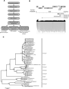
Figure 1
Figure 1
Application of DNA-capture-seq to analyze BLV proviral sequences and ISs. (A) Schematic diagram of the application of the target enrichment. (B) Visualization of sequence reads mapped to the FLK-BLV sequence (EF600696). NGS reads mapped to BLV are shown below the reference sequence. (C) Maximum-likelihood phylogenetic tree analysis of BLV whole genome sequences were generated with five newly obtained sequences (a sequence from FLK-BLV cell line indicated by filled square and four sequences from EBL tumor samples indicated by filled circle) together with 53 sequences from the GenBank database. The phylogenetic tree was generated and visualized using MEGA 7 with 1000 bootstrap replicates. The bar at the bottom of the figure denotes the estimated number of amino acid substitutions per site, indicating genetic variation for the length of the scale.
Figure 2
Figure 2
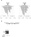
Visualization of ISs of the FLK-BLV cell line. (A) Visualization of NGS reads mapped to ovine genome around ISs. The arrow indicated the location of the IS. The sequences of virus-host chimeric reads next to the end of BLV gnome were indicated as host-left and host-right; respectively. (B) Confirmation of detected ISs by conventional PCR. M DNA ladder maker. Arrows indicate the positions of primers. ISs are numbered as in Table 1.
Figure 3
Figure 3
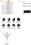
Visualization of ISs of EBL tumor, AS and PL blood samples. (A) Visualization of NGS reads mapped to the bovine genome around ISs. The arrow indicates the integration breakpoints in the bovine genome. (B) The pie-charts depict the distribution of clonal abundance of each EBL tumor sample, AS and PL blood sample. Each slice represents a BLV-infected clone and the size of the slice represents the relative abundance of the clone and the number indicates the proportion (%) of each clone. Oligoclonality index (OCI) measures the non-uniformity of the clonal abundance. ISs are numbered as in Table 2. (D) Visualization of NGS reads mapped to the bovine genome around the defective provirus of PL-2. The arrow indicates the integration breakpoints in the bovine genome.
Figure 4
Figure 4
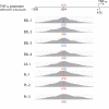
Amino acid polymorphisms of TNF-α promoter region. The sequence reads mapped to reference sequence (ARS-USD1.2/bosTau9) were visualized. Numbers denote the nucleotides count from the transcription initiation site. The nucleotides in the polymorphic sites are shown.

Similar articles

Cited by

References

    1. Barez PY, et al. Recent advances in BLV research. Viruses. 2015;7:6080–6088. doi: 10.3390/v7112929. - DOI - PMC - PubMed
    1. Gillet N, et al. Mechanisms of leukemogenesis induced by bovine leukemia virus: Prospects for novel anti-retroviral therapies in human. Retrovirology. 2007;4:18. doi: 10.1186/1742-4690-4-18. - DOI - PMC - PubMed
    1. Aida Y, Murakami H, Takahashi M, Takeshima SN. Mechanisms of pathogenesis induced by bovine leukemia virus as a model for human T-cell leukemia virus. Front. Microbiol. 2013;4:328. doi: 10.3389/fmicb.2013.00328. - DOI - PMC - PubMed
    1. Tsutsui T, Kobayashi S, Hayama Y, Yamamoto T. Fraction of bovine leukemia virus-infected dairy cattle developing enzootic bovine leukosis. Prev. Vet. Med. 2016;124:96–101. doi: 10.1016/j.prevetmed.2015.11.019. - DOI - PubMed
    1. Benitez OJ, Norby B, Bartlett PC, Maeroff JE, Grooms DL. Impact of bovine leukemia virus infection on beef cow longevity. Prev. Vet. Med. 2020;181:105055. doi: 10.1016/j.prevetmed.2020.105055. - DOI - PubMed

LinkOut - more resources