. 2021 Feb 25;11(1):4712.
doi: 10.1038/s41598-021-83825-6.

An NKX2-1GFP and TP63tdTomato dual fluorescent reporter for the investigation of human lung basal cell biology

Affiliations

An NKX2-1GFP and TP63tdTomato dual fluorescent reporter for the investigation of human lung basal cell biology

Kim Jee Goh et al. Sci Rep. .

Abstract

Basal cells are multipotent stem cells responsible for the repair and regeneration of all the epithelial cell types present in the proximal lung. In mice, the elusive origins of basal cells and their contribution to lung development were recently revealed by high-resolution, lineage tracing studies. It however remains unclear if human basal cells originate and participate in lung development in a similar fashion, particularly with mounting evidence for significant species-specific differences in this process. To address this outstanding question, in the last several years differentiation protocols incorporating human pluripotent stem cells (hPSC) have been developed to produce human basal cells in vitro with varying efficiencies. To facilitate this endeavour, we introduced tdTomato into the human TP63 gene, whose expression specifically labels basal cells, in the background of a previously described hPSC line harbouring an NKX2-1GFP reporter allele. The functionality and specificity of the NKX2-1GFP;TP63tdTomato hPSC line was validated by directed differentiation into lung progenitors as well as more specialised lung epithelial subtypes using an organoid platform. This dual fluorescent reporter hPSC line will be useful for tracking, isolating and expanding basal cells from heterogenous differentiation cultures for further study.


Conflict of interest statement

The authors declare no competing interests.

Figures

Figure 1
Figure 1
Schematic representation of the targeting strategy used to insert tdTomato into the endogenous TP63 locus in the NKX2-1GFP hiPSC reporter line. (a) Schematic of targeted NKX2-1 allele in BU3 NKX2-1GFP hiPSC line from Hawkins et al.. (b) Single guide RNAs were designed targeting the 3′ end of exon 14. Donor template consists of 800 bp homology arms flanking sequences encoding a P2A peptide (2A) and tdTomato. Expression of the α isoform of TP63 (both TAp63 and ΔNp63) is expected to lead to expression of tdTomato.
Figure 2
Figure 2
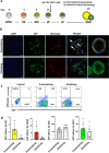
Assessment of tdTomato reporter functionality. (a) Schematic of lung differentiation protocol based on the work of McCauley et al. (2017). hPSC: human pluripotent stem cell; DE: definitive endoderm; AFE: anterior foregut endoderm; LP: lung progenitor. (b) Flow cytometry analysis of GFP + tdTomato + cells at Day 15 of the differentiation protocol. Representative dot plot shown. (c) Quantification of NKX2-1GFP+ cells at Day 15 as a measure of lung differentiation efficiency. Data are represented as means ± SD, n = 11 (NKX2-1GFP), n = 15 (NKX2-1GFP; TP63tdTomato). ns, not statistically significant. t test. Graph was made and statistical analysis was done using GraphPad Prism 8.0 (GraphPad Software Inc., San Diego, CA, USA) (www.graphpad.com).
Figure 3
Figure 3
Assessment of tdTomato reporter specificity. (a) QPCR analysis of TP63 (total), ∆Np63, TP63α and tdTomato expression in tdTomato- and tdTomato + fractions. n = 3. **p < 0.01, ***p < 0.001, ****p < 0.0001. t test. Graphs were made and statistical analyses were done using GraphPad Prism 8.0 (GraphPad Software Inc., San Diego, CA, USA) (www.graphpad.com). (b) Isolated tdTomato + cells plated onto Matrigel-coated plates and immunostained for TP63. Scale bar indicates 100 µm. (c) Western blot analysis of TP63 and Tubulin. 30 µg of whole cell lysates prepared from HEK293T cells overexpressing tdTomato (HEK293T), TP63TOM + cells, and primary human bronchial epithelial cells (priHBEC). Blots were probed with anti-TP63 antibody with Tubulin used as a loading control. Bands corresponding to TP63 and Tubulin are indicated by black arrows. The asterisk indicates where the uncleaved TP63-P2A-tdTomato protein of about 110 kDa is expected to be, if present. The full uncropped blots are presented in Supplementary Fig. 5.
Figure 4
Figure 4
Differentiation of NKX2-1GFP;TP63tdTomato hiPSCs into lung organoids. (a) Protocol for differentiating NKX2-1GFP;TP63tdTomato hiPSCs into proximalized and distalized lung organoids based on McCauley et al. and Jacob et al.. hPSC human pluripotent stem cell, DE definitive endoderm, AFE anterior foregut endoderm, LP lung progenitors, ORG lung organoid. (b) Representative images of proximalized and distalized lung organoids derived from the NKX2-1GFP;TP63tdTomato hiPSC line. White arrows indicate lung organoids containing double positive cells. (c) Flow cytometry analysis of lung organoids cultured in proximalizing and distalizing conditions. Representative dot plots shown. (d) Quantification of the 4 populations in the lung organoids grown in the differentiation culture conditions. DN double negative (GFP- tdTOM-), DP double positive (GFP + tdTOM +), tdTomato + tdTomato-positive only (GFP- tdTOM +), GFP GFP-positive only (GFP + tdTomato-). Data are represented as means ± SD, n = 5. *p < 0.05. t test. Graphs were made and statistical analyses were done using GraphPad Prism 8.0 (GraphPad Software Inc., San Diego, CA, USA) (www.graphpad.com).
Figure 5
Figure 5
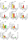
Gene expression analysis of NKX2-1GFP;TP63tdTomato hiPSC-derived lung organoids. (a) QPCR analysis of lung marker NKX2-1 and basal cell markers TP63 (total), ΔNp63, TP63α, KRT5, and NGFR. Expression levels were normalized to housekeeping gene, ACTB. n = 3. *p < 0.05. t test. (b) QPCR analysis of additional markers identified by Miller et al. (2020) to be associated with basal cells. Expression levels were normalized to ACTB. n = 3. **p < 0.01. All graphs were made and statistical analyses were done using GraphPad Prism 8.0 (GraphPad Software Inc., San Diego, CA, USA) (www.graphpad.com).
Figure 6
Figure 6
Analysis of basal cell marker expression in 4 cell fractions isolated from Day 30 proximalized lung organoids. (a) QPCR analysis of basal cell markers in DN, DP, tdTomato + and GFP + fractions isolated from Day 30 proximalized lung organoids. Expression levels were normalized to ACTB. DN double negative (GFP- tdTOM-), DP double positive (GFP + tdTOM +), tdTomato +  tdTomato-positive only (GFP- tdTOM +), GFP GFP-positive only (GFP + tdTomato-), hiPSC undifferentiated BU3 NKX2-1GFP hiPSCs. n = 4. **p < 0.01, ***p < 0.001, ****p < 0.0001. ANOVA followed by Tukey’s posthoc test. (b) QPCR analysis of other basal cell markers EGFR, F3, S100A2 and IL-33 in the DN, DP, tdTomato + and GFP + fractions. Expression levels were normalized to ACTB. n = 4. **p < 0.01, ***p < 0.001, ****p < 0.0001. ANOVA followed by Tukey’s post hoc test. All graphs were made and statistical analyses were done using GraphPad Prism 8.0 (GraphPad Software Inc., San Diego, CA, USA) (www.graphpad.com).
Figure 7
Figure 7
DP cells are proliferative and are capable of self-renewal. (a) Schematic of experimental setup. DP cells were serially passaged up to 3 passages. ORG: lung organoid; DP: double positive; MG: Matrigel. (b) Growth kinetics of DP cells at passage number 1 (P1). n = 3. (c) DP cells quantified at passage numbers P0, P1, P2 and P3 using flow cytometry. n = 3. (b,c) were made and statistical analyses were done using GraphPad Prism 8.0 (GraphPad Software Inc., San Diego, CA, USA) (www.graphpad.com). (d) Representative images of organoids derived from DP cells at P3. Scale bars indicate 500 µm. (e) Representative flow cytometry dot plot of dissociated organoids derived from DP P3 cells.
Figure 8
Figure 8
DP cells possess multilineage differentiation potential. (a) Schematic of differentiation protocol. Isolated DP cells were seeded onto transwells and then cultured at an air–liquid interface (ALI) upon reaching confluence in the absence or presence of 10 ng/ml IL-6 or 10 ng/ml IL-13. (b) Representative images of differentiation cultures immunostained for acetylated α-tubulin (AcTUB) and MUC5AC. Scale bars indicate 5 µm. n = 3. (c) QPCR analysis of multiciliated cell markers (FOXJ1, DNAH5, SNTN), secretory cell markers (SPDEF, MUC5AC, MUC5B). n = 3. Graphs were made and statistical analyses were done using GraphPad Prism 8.0 (GraphPad Software Inc., San Diego, CA, USA) (www.graphpad.com).

Similar articles

Cited by

References

    1. Society, E. R. Forum of International Respiratory Societies. The Global Impact of Respiratory Disease – Second Edition. Sheffield,. (2017).
    1. Bowden DH. Cell turnover in the lung. Am. Rev. Respir. Dis. 1983;128:2. - PubMed
    1. Kauffman SL. Cell proliferation in the mammalian lung. Int. Rev. Exp. Pathol. 1980;22:131–191. - PubMed
    1. Adamson IYR, Bowden DH. The type 2 cell as progenitor of alveolar epithelial regeneration: A cytodynamic study in mice after exposure to oxygen. Lab. Investig. 1974;30:35–42. - PubMed
    1. Adamson IYR, Bowden DH. Origin of ciliated alveolar epithelial cells in bleomycin induced lung injury. Am. J. Pathol. 1977;87:569–580. - PMC - PubMed

Publication types

MeSH terms

LinkOut - more resources