

A novel technology to integrate imaging and clinical markers for non-invasive diagnosis of lung cancer
- PMID: 33633213
- PMCID: PMC7907202
- DOI: 10.1038/s41598-021-83907-5
A novel technology to integrate imaging and clinical markers for non-invasive diagnosis of lung cancer
Abstract
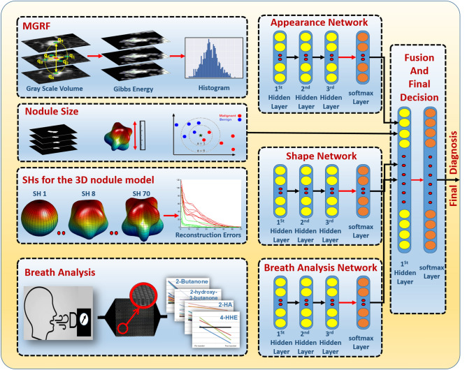
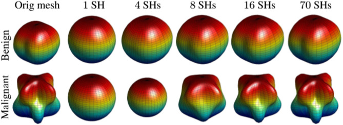
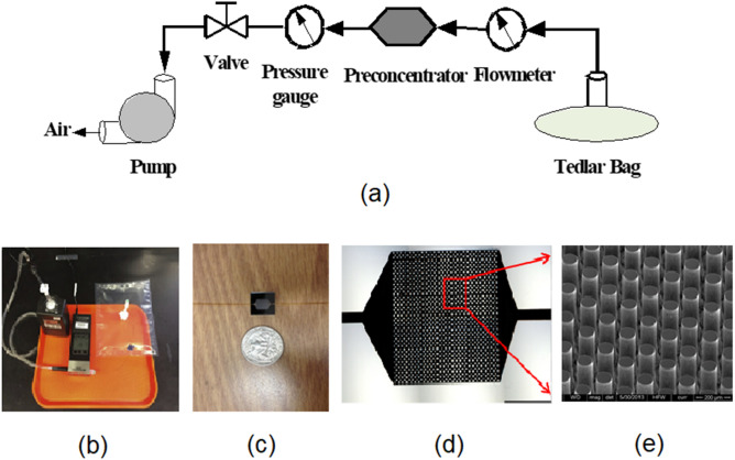
This study presents a non-invasive, automated, clinical diagnostic system for early diagnosis of lung cancer that integrates imaging data from a single computed tomography scan and breath bio-markers obtained from a single exhaled breath to quickly and accurately classify lung nodules. CT imaging and breath volatile organic compounds data were collected from 47 patients. Spherical Harmonics-based shape features to quantify the shape complexity of the pulmonary nodules, 7th-Order Markov Gibbs Random Field based appearance model to describe the spatial non-homogeneities in the pulmonary nodule, and volumetric features (size) of pulmonary nodules were calculated from CT images. 27 VOCs in exhaled breath were captured by a micro-reactor approach and quantied using mass spectrometry. CT and breath markers were input into a deep-learning autoencoder classifier with a leave-one-subject-out cross validation for nodule classification. To mitigate the limitation of a small sample size and validate the methodology for individual markers, retrospective CT scans from 467 patients with 727 pulmonary nodules, and breath samples from 504 patients were analyzed. The CAD system achieved 97.8% accuracy, 97.3% sensitivity, 100% specificity, and 99.1% area under curve in classifying pulmonary nodules.
Conflict of interest statement
The authors declare no competing interests.
Figures

Similar articles
-
A volatile biomarker in breath predicts lung cancer and pulmonary nodules.J Breath Res. 2019 Jun 19;13(3):036013. doi: 10.1088/1752-7163/ab21aa. J Breath Res. 2019. PMID: 31085817
-
Differentiating Pulmonary Nodule Malignancy Using Exhaled Volatile Organic Compounds: A Prospective Observational Study.Cancer Med. 2025 Jan;14(1):e70545. doi: 10.1002/cam4.70545. Cancer Med. 2025. PMID: 39777868 Free PMC article.
-
Breath carbonyl compounds as biomarkers of lung cancer.Lung Cancer. 2015 Oct;90(1):92-7. doi: 10.1016/j.lungcan.2015.07.005. Epub 2015 Jul 19. Lung Cancer. 2015. PMID: 26233567
-
Inconsistencies in predictive models based on exhaled volatile organic compounds for distinguishing between benign pulmonary nodules and lung cancer: a systematic review.BMC Pulm Med. 2024 Nov 2;24(1):551. doi: 10.1186/s12890-024-03374-2. BMC Pulm Med. 2024. PMID: 39488679 Free PMC article.
-
Lung cancer biomarkers in exhaled breath.Expert Rev Mol Diagn. 2011 Mar;11(2):207-17. doi: 10.1586/erm.10.112. Expert Rev Mol Diagn. 2011. PMID: 21405971 Review.
Cited by
-
Minimally invasive biomarkers for triaging lung nodules-challenges and future perspectives.Cancer Metastasis Rev. 2025 Jan 31;44(1):29. doi: 10.1007/s10555-025-10247-5. Cancer Metastasis Rev. 2025. PMID: 39888565 Free PMC article. Review.
-
Dynamic learning for imbalanced data in learning chest X-ray and CT images.Heliyon. 2023 Jun 1;9(6):e16807. doi: 10.1016/j.heliyon.2023.e16807. eCollection 2023 Jun. Heliyon. 2023. PMID: 37313141 Free PMC article.
-
[Advances on Collection and Analysis of Volatile Organic Compounds in the Diagnosis of Lung Cancer].Zhongguo Fei Ai Za Zhi. 2021 Nov 20;24(11):796-803. doi: 10.3779/j.issn.1009-3419.2021.101.41. Zhongguo Fei Ai Za Zhi. 2021. PMID: 34802212 Free PMC article. Chinese.
-
Chest CT signs and serum homocysteine levels can effectively diagnose chronic heart failure.Am J Transl Res. 2025 Apr 15;17(4):3085-3093. doi: 10.62347/BOIQ3903. eCollection 2025. Am J Transl Res. 2025. PMID: 40385067 Free PMC article.
-
Deep learning facilitates multi-data type analysis and predictive biomarker discovery in cancer precision medicine.Comput Struct Biotechnol J. 2023 Jan 31;21:1372-1382. doi: 10.1016/j.csbj.2023.01.043. eCollection 2023. Comput Struct Biotechnol J. 2023. PMID: 36817954 Free PMC article. Review.
References
-
- American Cancer Society . Cancer Facts and Figures. Providence: American Cancer Society; 2019.
-
- Ries, L. A. G. et al. Cancer survival among adults: Us seer program, 1988–2001. Patient and tumor characteristics SEER Survival Monograph Publication07–6215 (2007).
MeSH terms
Substances
LinkOut - more resources
Full Text Sources
Other Literature Sources
Medical
Miscellaneous
