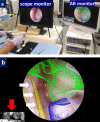
Experimental pilot study for augmented reality-enhanced elbow arthroscopy
- PMID: 33633227
- PMCID: PMC7907139
- DOI: 10.1038/s41598-021-84062-7
Experimental pilot study for augmented reality-enhanced elbow arthroscopy
Erratum in
-
Author Correction: Experimental pilot study for augmented reality-enhanced elbow arthroscopy.Sci Rep. 2021 May 17;11(1):10708. doi: 10.1038/s41598-021-90073-1. Sci Rep. 2021. PMID: 34001995 Free PMC article. No abstract available.
Abstract
The purpose of this study was to develop and evaluate a novel elbow arthroscopy system with superimposed bone and nerve visualization using preoperative computed tomography (CT) and magnetic resonance imaging (MRI) data. We obtained bone and nerve segmentation data by CT and MRI, respectively, of the elbow of a healthy human volunteer and cadaveric Japanese monkey. A life size 3-dimensional (3D) model of human organs and frame was constructed using a stereo-lithographic 3D printer. Elbow arthroscopy was performed using the elbow of a cadaveric Japanese monkey. The augmented reality (AR) range of error during rotation of arthroscopy was examined at 20 mm scope-object distances. We successfully performed AR arthroscopy using the life-size 3D elbow model and the elbow of the cadaveric Japanese monkey by making anteromedial and posterior portals. The target registration error was 1.63 ± 0.49 mm (range 1-2.7 mm) with respect to the rotation angle of the lens cylinder from 40° to - 40°. We attained reasonable accuracy and demonstrated the operation of the designed system. Given the multiple applications of AR-enhanced arthroscopic visualization, it has the potential to be a next-generation technology for arthroscopy. This technique will contribute to the reduction of serious complications associated with elbow arthroscopy.
Conflict of interest statement
The authors declare no competing interests.
Figures


Similar articles
-
Augmented reality-based surgical guidance for wrist arthroscopy with bone-shift compensation.Comput Methods Programs Biomed. 2023 Mar;230:107323. doi: 10.1016/j.cmpb.2022.107323. Epub 2022 Dec 23. Comput Methods Programs Biomed. 2023. PMID: 36608430
-
Elbow arthroscopy: An alternative to anteromedial portals.Orthop Traumatol Surg Res. 2015 Jun;101(4):411-4. doi: 10.1016/j.otsr.2015.03.011. Epub 2015 Apr 21. Orthop Traumatol Surg Res. 2015. PMID: 25910702
-
Total elbow arthroplasty using an augmented reality-assisted surgical technique.J Shoulder Elbow Surg. 2022 Jan;31(1):175-184. doi: 10.1016/j.jse.2021.05.019. Epub 2021 Jun 25. J Shoulder Elbow Surg. 2022. PMID: 34175467
-
Safety of Anteromedial Portals in Elbow Arthroscopy: A Systematic Review of Cadaveric Studies.Arthroscopy. 2019 Jul;35(7):2164-2172. doi: 10.1016/j.arthro.2019.02.046. Arthroscopy. 2019. PMID: 31272638 Free PMC article.
-
[Extra-articular elbow arthroscopy].Chir Main. 2006 Nov;25 Suppl 1:S121-30. Chir Main. 2006. PMID: 17361882 Review. French.
Cited by
-
Eyes and movement differences in unconscious state during microscopic procedures.Sci Rep. 2025 Feb 25;15(1):6712. doi: 10.1038/s41598-025-88647-4. Sci Rep. 2025. PMID: 40000779 Free PMC article.
-
Accessibility of osteochondral lesion at the capitellum during elbow arthroscopy: an anatomical study.Arch Orthop Trauma Surg. 2024 Mar;144(3):1297-1302. doi: 10.1007/s00402-023-05172-7. Epub 2024 Jan 3. Arch Orthop Trauma Surg. 2024. PMID: 38172435 Free PMC article.
-
Validation Study on Iatrogenic Nerve Damage Reduction Using Augmented Reality on Elbow Phantom.Mayo Clin Proc Digit Health. 2025 Apr 16;3(2):100221. doi: 10.1016/j.mcpdig.2025.100221. eCollection 2025 Jun. Mayo Clin Proc Digit Health. 2025. PMID: 40487861 Free PMC article.
-
Intra-Articular Ultrasonography Probe for Minimally Invasive Upper Extremity Arthroscopic Surgery: A Phantom Study.J Clin Med. 2023 Sep 2;12(17):5727. doi: 10.3390/jcm12175727. J Clin Med. 2023. PMID: 37685794 Free PMC article.
References
Publication types
MeSH terms
LinkOut - more resources
Full Text Sources
Other Literature Sources
Research Materials

