

Association of changes of retinal vessels diameter with ocular blood flow in eyes with diabetic retinopathy
- PMID: 33633255
- PMCID: PMC7907275
- DOI: 10.1038/s41598-021-84067-2
Association of changes of retinal vessels diameter with ocular blood flow in eyes with diabetic retinopathy
Abstract
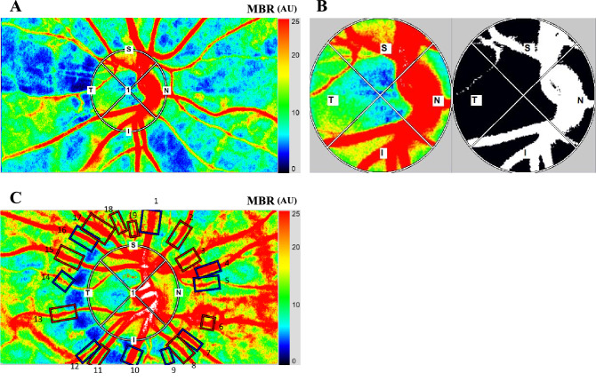
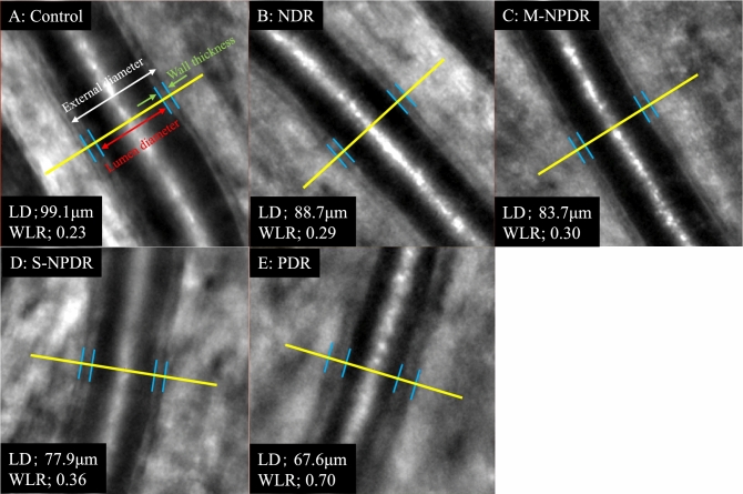
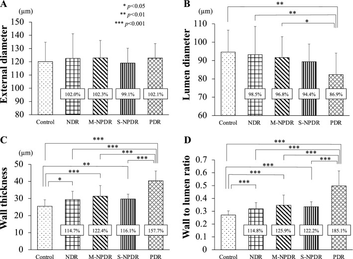
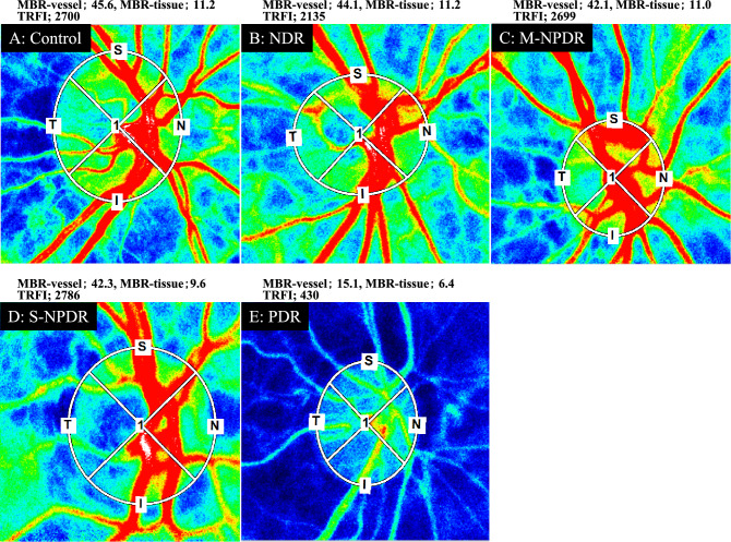
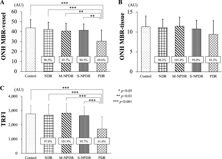
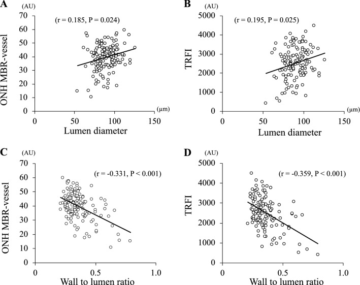
We investigated morphological changes of retinal arteries to determine their association with the blood flow and systemic variables in type 2 diabetes patients. The patients included 47 non-diabetic retinopathy eyes, 36 mild or moderate nonproliferative diabetic retinopathy (M-NPDR) eyes, 22 severe NPDR (S-NPDR) eyes, 32 PDR eyes, and 24 normal eyes as controls. The mean wall to lumen ratio (WLR) measured by adaptive optics camera was significantly higher in the PDR groups than in all of the other groups (all P < 0.001). However, the external diameter of the retinal vessels was not significantly different among the groups. The mean blur rate (MBR)-vessel determined by laser speckle flowgraphy was significantly lower in the PDR group than in the other groups (P < 0.001). The WLR was correlated with MBR-vessel (r = - 0.337, P < 0.001), duration of disease (r = 0.191, P = 0.042), stage of DM (r = 0.643, P < 0.001), systolic blood pressure (r = 0.166, P < 0.037), and presence of systemic hypertension (r = 0.443, P < 0.001). Multiple regression analysis demonstrated that MBR-vessel (β = - 0.389, P < 0.001), presence of systemic hypertension (β = 0.334, P = 0.001), and LDL (β = 0.199, P = 0.045) were independent factors significantly associated with the WLR. The increased retinal vessel wall thickness led to a narrowing of lumen diameter and a decrease in the blood flow in the PDR group.
Conflict of interest statement
The authors declare no competing interests.
Figures
Similar articles
-
Effects of photocoagulation on ocular blood flow in patients with severe non-proliferative diabetic retinopathy.PLoS One. 2017 Mar 29;12(3):e0174427. doi: 10.1371/journal.pone.0174427. eCollection 2017. PLoS One. 2017. PMID: 28355247 Free PMC article.
-
Interaction Between the Distribution of Diabetic Retinopathy Lesions and the Association of Optical Coherence Tomography Angiography Scans With Diabetic Retinopathy Severity.JAMA Ophthalmol. 2020 Dec 1;138(12):1291-1297. doi: 10.1001/jamaophthalmol.2020.4516. JAMA Ophthalmol. 2020. PMID: 33119083 Free PMC article.
-
Evaluation of retinal blood flow before and after panretinal photocoagulation using pattern scan laser for diabetic retinopathy.Curr Eye Res. 2017 Dec;42(12):1707-1712. doi: 10.1080/02713683.2017.1358373. Epub 2017 Sep 22. Curr Eye Res. 2017. PMID: 28937857
-
QUANTIFICATION OF RETINAL VESSEL TORTUOSITY IN DIABETIC RETINOPATHY USING OPTICAL COHERENCE TOMOGRAPHY ANGIOGRAPHY.Retina. 2018 May;38(5):976-985. doi: 10.1097/IAE.0000000000001618. Retina. 2018. PMID: 28333883
-
Review and comparison of retinal vessel calibre and geometry software and their application to diabetes, cardiovascular disease, and dementia.Graefes Arch Clin Exp Ophthalmol. 2023 Aug;261(8):2117-2133. doi: 10.1007/s00417-023-06002-7. Epub 2023 Feb 18. Graefes Arch Clin Exp Ophthalmol. 2023. PMID: 36801971 Review.
Cited by
-
Evaluation of the Effect of Garlic Tablet as a Complementary Treatment for Patients with Diabetic Retinopathy.J Diabetes Res. 2022 Jul 14;2022:6620661. doi: 10.1155/2022/6620661. eCollection 2022. J Diabetes Res. 2022. PMID: 35875346 Free PMC article. Clinical Trial.
-
Multimodal Imaging of Diabetic Retinopathy: Insights from Optical Coherence Tomography Angiography and Adaptive Optics.Diagnostics (Basel). 2025 Jul 8;15(14):1732. doi: 10.3390/diagnostics15141732. Diagnostics (Basel). 2025. PMID: 40722482 Free PMC article.
-
Adaptive vessel tracing and segmentation in OCT enables the robust detection of wall-to-lumen ratio abnormalities in 5xFAD mice.Biomed Opt Express. 2023 Nov 20;14(12):6350-6360. doi: 10.1364/BOE.504317. eCollection 2023 Dec 1. Biomed Opt Express. 2023. PMID: 38420326 Free PMC article.
-
Hemodynamic and morphological changes of the central retinal artery in myopic eyes.Sci Rep. 2022 May 2;12(1):7104. doi: 10.1038/s41598-022-11087-x. Sci Rep. 2022. PMID: 35501327 Free PMC article.
-
Combined optical coherence tomography and electroretinography system for imaging neurovascular coupling in the human retina.Neurophotonics. 2025 Jul;12(3):035004. doi: 10.1117/1.NPh.12.3.035004. Epub 2025 Aug 9. Neurophotonics. 2025. PMID: 40786701 Free PMC article.
References
MeSH terms
LinkOut - more resources
Full Text Sources
Other Literature Sources
Medical
