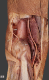
Impact of plantaris ligamentous tendon
- PMID: 33633305
- PMCID: PMC7907062
- DOI: 10.1038/s41598-021-84186-w
Impact of plantaris ligamentous tendon
Abstract
There are countless morphological variations among the muscles, tendons, ligaments, arteries, veins and nerves of the human body, many of which remain undescribed. Anatomical structures are also subject to evolution, many disappearing and others continually emerging. The main goal of this pilot study was to describe a previously undetected anatomical structure, the plantaris ligamentous tendon, and to determine its frequency and histology. Twenty-two lower limbs from 11 adult cadavers (11 left, and 11 right) fixed in 10% formalin were examined. The mean age of the cadavers at death was 60.1 years (range 38-85). The group comprised six women and five men from a Central European population. All anatomical dissections of the leg and foot area accorded with the pre-established protocol. Among the 22 lower limbs, the PLT was present in 16 (72.7%) and absent in six (27.3%). It originated as a strong fan-shaped ligamentous tendon from the superior part of the plantaris muscle, the posterior surface of the femur and the lateral aspect of the knee joint capsule. It inserted to the ilio-tibial band. Histologically, a tendon and ligament were observed extending parallel to each other. A new anatomical structure has been found, for which the name plantaris ligamentous tendon is proposed. It occurs around the popliteal region between the plantaris muscle, the posterior surface of the femur, and the ilio-tibial band.
Conflict of interest statement
The authors declare no competing interests.
Figures


Similar articles
-
Proposal for a new classification of plantaris muscle origin and its potential effect on the knee joint.Ann Anat. 2020 Sep;231:151506. doi: 10.1016/j.aanat.2020.151506. Epub 2020 Mar 12. Ann Anat. 2020. PMID: 32173563
-
Never undescribed before, four-headed plantaris muscle.Folia Morphol (Warsz). 2024;83(4):942-946. doi: 10.5603/fm.98753. Epub 2024 May 17. Folia Morphol (Warsz). 2024. PMID: 38757503
-
Possible effect of morphological variations of plantaris muscle tendon on harvesting at reconstruction surgery-case report.Surg Radiol Anat. 2020 Oct;42(10):1183-1188. doi: 10.1007/s00276-020-02463-1. Epub 2020 Apr 4. Surg Radiol Anat. 2020. PMID: 32248255 Free PMC article.
-
The plantaris muscle: too important to be forgotten. A review of evolution, anatomy, clinical implications and biomechanical properties.J Sports Med Phys Fitness. 2019 May;59(5):839-845. doi: 10.23736/S0022-4707.18.08816-3. J Sports Med Phys Fitness. 2019. PMID: 30936418 Review.
-
A Meta-Analysis of the Surgical Availability and Morphology of the Plantaris Tendon.J Hand Surg Asian Pac Vol. 2019 Jun;24(2):208-218. doi: 10.1142/S2424835519500280. J Hand Surg Asian Pac Vol. 2019. PMID: 31035870 Review.
Cited by
-
Relationships among Coracobrachialis, Biceps Brachii, and Pectoralis Minor Muscles and Their Correlation with Bifurcated Coracoid Process.Biomed Res Int. 2022 Mar 25;2022:8939359. doi: 10.1155/2022/8939359. eCollection 2022. Biomed Res Int. 2022. PMID: 35419460 Free PMC article.
-
The accessory heads of the quadriceps femoris muscle may affect the layering of the quadriceps tendon and potential graft harvest lengths.Knee Surg Sports Traumatol Arthrosc. 2023 Dec;31(12):5755-5764. doi: 10.1007/s00167-023-07647-x. Epub 2023 Nov 6. Knee Surg Sports Traumatol Arthrosc. 2023. PMID: 37932536 Free PMC article.
-
The Plantaris Muscle Is Not Vestigial: Developmental, Comparative, and Functional Evidence for Its Sensorimotor Role.Biology (Basel). 2025 Jun 13;14(6):696. doi: 10.3390/biology14060696. Biology (Basel). 2025. PMID: 40563947 Free PMC article. Review.
-
Unknown variant of the accessory subscapularis muscle?Anat Sci Int. 2022 Jan;97(1):138-142. doi: 10.1007/s12565-021-00633-8. Epub 2021 Sep 30. Anat Sci Int. 2022. PMID: 34591277 Free PMC article.
-
Does the Anatomical Type of the Plantaris Tendon Influence the Management of Midportion Achilles Tendinopathy?J Clin Med. 2025 Aug 4;14(15):5478. doi: 10.3390/jcm14155478. J Clin Med. 2025. PMID: 40807104 Free PMC article. Review.
References
MeSH terms
LinkOut - more resources
Full Text Sources
Other Literature Sources

