. 2021 Mar;28(3):278-289.
doi: 10.1038/s41594-021-00560-2. Epub 2021 Feb 25.

Target-induced clustering activates Trim-Away of pathogens and proteins

Affiliations

Target-induced clustering activates Trim-Away of pathogens and proteins

Jingwei Zeng et al. Nat Struct Mol Biol. 2021 Mar.

Abstract

Trim-Away is a recently developed technology that exploits off-the-shelf antibodies and the RING E3 ligase and cytosolic antibody receptor TRIM21 to carry out rapid protein depletion. How TRIM21 is catalytically activated upon target engagement, either during its normal immune function or when repurposed for targeted protein degradation, is unknown. Here we show that a mechanism of target-induced clustering triggers intermolecular dimerization of the RING domain to switch on the ubiquitination activity of TRIM21 and induce virus neutralization or drive Trim-Away. We harness this mechanism for selective degradation of disease-causing huntingtin protein containing long polyglutamine tracts and expand the Trim-Away toolbox with highly active TRIM21-nanobody chimeras that can also be controlled optogenetically. This work provides a mechanism for cellular activation of TRIM RING ligases and has implications for targeted protein degradation technologies.


Conflict of interest statement

Competing Interests

The authors declare no competing interests.

Figures
Extended Data Fig. 1
Extended Data Fig. 2
Extended Data Fig. 3
Extended Data Fig. 4
Extended Data Fig. 5
Fig. 1
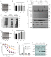
Fig. 1. RING dimerization is required for TRIM21 activation
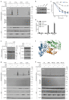
a, Crystal structure of TRIM21 RING dimer (PDB 5OLM) showing the important residues M10 and M72 at the dimer interface. b, Immunoblots showing Trim-away of target protein IKKα following electroporation (+) of anti-IKKα IgG (anti-IKKα). IKKα degradation is deficient in cells expressing the M72E mutant TRIM21. c–e, T21RLR shows enhanced ubiquitination chain formation (c) and ubiquitin discharge from Ube2N~Ub (d,e) compared to T21R. Immunoblots in b,c,e are representative examples from three independent experiments. f, Top, dummy atom model (DAM) structures calculated from SAXS data (Supplementary Note 1) for T21RBCC (purple spheres) and T21CC235 (blue spheres). Bottom, structural model of T21RBCC based on atomic models of the TRIM21 RING (yellow) and B Box (orange) domains (PDB 5OLM) and TRIM25 coiled-coil domain (cyan, PDB 4CFG). g, Averaged and filtered DAM’s (white and green spheres, respectively) from TRIM21:Fc SAXS data (Supplementary Note 1) overlaid with an atomic model of the TRIM21:Fc complex using PDB 5OLM (RING, B Box), PDB 4CFG (coiled-coil) and PDB 2IWG (PRYSPRY (magenta) and IgG Fc (grey)). h, Schematic of potential intermolecular RING dimerisation on the surface of a virus-antibody complex. i, Remaining infectivity of AdV5 following incubation with saturating concentrations of varying ratios of WT:H433A 9C12 antibody. j, Neutralisation of AdV5 infection by increasing 9C12 concentrations in cells expressing the indicated TRIM21 mutants. k, AdV5-9C12-induced NFkB activation in cells expressing the indicated TRIM21 mutants. Graphs in panels d,i–k show mean and s.e.m. for n=3 independent experiments; Statistical significance is based on Student’s t-test (d), two-way ANOVA (j) and one-way ANOVA (k) and represented with labels ns (not significant, P>0.05), **(P≤0.01), ****(P≤0.0001), as defined in Methods. Source data for graphs and statistics are in Supplementary Data 1. Uncropped blots are shown in Supplementary Data 2.
Fig. 2
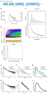
Fig. 2. Trim-Away requires recruitment of multiple TRIM21 molecules to the target.
a, Schematic of myc-mEGFP constructs. b-d, HEK293T-mCherry-TRIM21 cells were electroporated with mRNA encoding the indicated myc-mEGFP constructs together with either control IgG (9C12), ant-myc (9E10) or anti-GFP (polyclonal) antibodies. 8h post-electroporation cellular GFP fluorescence was imaged (b) and quantified (c) using the IncuCyte system, or total GFP protein levels analysed by immunoblotting (d). Scale bars, 100 μm. e, HEK293T-mCherry-TRIM21 cells were electroporated with mRNA encoding the indicated myc-mEGFP constructs together with control IgG or increasing concentrations of anti-myc antibody and GFP fluorescence quantified 8 hours later using the IncuCyte. f, HEK293T-mCherry-TRIM21 cells were electroporated with mRNA encoding 2myc-mEGFP together with the indicated antibodies and GFP fluorescence quantified 8 hours later using the IncuCyte. g-–i The indicated antibodies (50 nM) either alone, or mixed with GFP protein (100 nM), were analysed by mass photometry. j, RPE-1 cells expressing mEGFP were electroporated with the indicated antibodies and cell extracts immunoblotted 3 hours later. Incucyte images (b), Immunoblots (d,j) and mass photometry profiles (g-i) are representative examples from three independent experiments. Graphs in c,e,f show mean and s.e.m. (some error bars are to small to be seen) from n=3 independent experiments. Statistical significance was calculated based on two-tailed Student’s t-test and represented with labels ns (not significant, P>0.05), *** (P≤0.001), ****(P≤0.0001), as defined in Methods. Source data for graphs and statistics are in Supplementary Data 1. Uncropped blots are in Supplementary Data 2.
Fig. 3
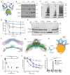
Fig. 3. The nature of the target dictates TRIM21 activation
a, mEGFP is a monomer and exhibits diffuse localisation. b,c, H2B-mEGFP and mEGFP-PACT are incorporated into histones and centrosomes respectively, which act as oligomeric scaffolds for mEGFP. di, RPE-1 cells were electroporated with mRNA encoding mEGFP (d,e), H2B-mEGFP (f,g) or mEGFP-PACT (h,i) alone or together with vhhGFP4-Fc or vhhGFP4-Fc(H433A) and fixed 16h later for immunofluorescence. Representative confocal microscope images (d,f,h) and quantification (e,g,i) show that mEGFP is degraded in a TRIM21-dependent manner by vhhGFP4-Fc only when tethered to the oligomeric histone and centrosome scaffolds. Scale bars, 10 μm. Yellow arrows in h show centrosomes. Graphs in e,g,i show mean and s.d. from n=3 independent experiments. Number of cells quantified in brackets. P values from two-tailed Student’s t-test. j, Schematic of mEGFP fused to oligomeric Caveolin-1. k,i, WT NIH3T3 or NIH3T3-Caveolin-1-GFP cells were electroporated with mRNA encoding vhhGFP4-Fc or vhhGFP4-Fc(H433A) and 16 hours later analysed by immunoblotting (k) and confocal microscopy (l). Scale bars, 50 μm. Source data for graphs and statistics are in Supplementary Data 1.
Fig. 4
Fig. 4. Selective degradation of disease-causing huntingtin by TRIM21
a, Schematic of human huntingtin protein highlighting the N-terminal polyglutamine (polyQ) tract. PolyQ tracts greater than 39Q are associated with Huntington’s disease. The anti-polyQ monoclonal antibody 3B5H10 binds to a 13Q epitope. Up to two 3B5H10 antibodies are predicted to bind to normal huntingtin, with additional antibodies predicted to bind as the length of polyQ increases, which would lead to TRIM21 clustering, activation and degradation of only huntingtin protein with long, disease-causing, polyQ tracts. b,c, HEK293T-mCherry-TRIM21 were electroporated with mRNA encoding the indicated polyQ-mEGFP constructs together with either anti-polyQ (3B5H10), control IgG (9C12) or anti-GFP (polyclonal) antibodies. 8 hours post-electroporation cellular GFP fluorescence was imaged (b) and quantified (c) using the IncuCyte system. Scale bars 100 μm. Data points in c show mean and s.e.m. (some error bars are too small to be seen), n=3 independent experiments. Statistical significance is based on two-tailed Student’s t-test and represented with labels: ns (not significant, P>0.05), **(P ≤ 0.01), ****(P ≤ 0.0001), as defined in Methods. Source data for graphs and statistics are in Supplementary Data 1.
Fig. 5
Fig. 5. Molecular clustering activates TRIM21
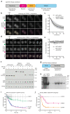
a, Schematic of light-induced clustering of TRIM21. b,c, Drosophila S2 cells expressing the indicated constructs were incubated with or without MG132 and RFP fluorescence quantified by live imaging. Time shows minutes (min) from onset of blue light exposure. Scale bars, 5 μm. Pseudo-coloured kymographs show fluorescence intensity in regions defined by red dotted lines. Graph shows mean and s.d. of fluorescence intensity of RFP-CRY2-TRIM21 (n=23 cells) and RFP-CRY2-TRIM21+MG132 (n=20 cells) normalised for the respective controls (RFP-CRY2 (n=16 cells) and RFP-CRY2 + MG132 (n=15 cells). d, RPE-1 cells expressing the indicated constructs together with mem-mEGFP were incubated with or without MG132 and blue light for 3 hours prior to immunoblotting. Representative immunoblots from three independent experiments. Source data for graphs are Supplementary Data 1. Uncropped blots are in Supplementary Data 2.
Fig. 6
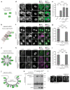
Fig. 6. New ways to Trim-Away proteins
a, Schematic of optoTrim-Away construct. b-e, Drosophila S2 cells expressing GFP-aPKC and vhhGFP4-RFP-CRY2-TRIM21 (b,c) or RFP-CRY2-vhhGFP4 (d,e) were analysed by live imaging. Time shows minutes (min) from onset of blue light exposure. Scale bars, 10 μm. Graphs show mean and s.d. of GFP and RFP fluorescence intensity (n=30 cells in c and n=24 cells in e). f, RPE-1 cells expressing the indicated constructs together with mem-mEGFP were incubated with or without MG132 and blue light for 3 hours prior to immunoblotting. g, Schematic of miniTrim-Away construct. h, Bacterially-expressed 6His-T21R-vhhGFP4 was purified using a two-step protocol of NiNTA- followed by size exclusion- chromatography and analysed by Coomassie blue staining of SDS-PAGE. i, NIH3T3-Caveolin-1-GFP cells were electroporated with PBS (control) or the indicated concentrations of T21R-vhhGFP4 protein and GFP fluorescence was quantified using the IncuCyte system. j, H2B-GFP primary MEFs were electroporated with PBS (control) or T21R-vhhGFP4 in the form or mRNA or protein and GFP fluorescence quantified using the IncuCyte system. Graphs in i and j show mean and s.e.m. of n=4 technical replicates. Representative immunoblots (f) and GFP degradation profiles (i,j) from three independent experiments. Source data for graphs are Supplementary Data 1. Uncropped blots are in Supplementary Data 2.
Fig. 7
Fig. 7. Model for target-induced clustering mechanism and its applications
The two RING domains of the TRIM21 dimer are held apart by an elongated antiparallel coiled-coil. Target-induced clustering allows intermolecular RING dimerisation, which facilitates E2~Ub binding and catalysis of K63-linked ubiquitin chains on TRIM21 leading to proteasomal degradation of TRIM21 and its targets. TRIM21 clustering can be induced by antibody-coated virus, internalised immune complexes or proteins targeted with antibodies. This mechanism has therapeutic potential for selective degradation of repeat-epitope or aggregated disease proteins (orange shaded area). New protein depletion tools based on the sufficiency of molecular clustering and RING dimerisation for TRIM21-mediated degradation (green shaded area).

Similar articles

Cited by

References

    1. Zheng N, Shabek N. Ubiquitin Ligases: Structure, Function, and Regulation. Annu Rev Biochem. 2017;86:129–157. - PubMed
    1. Schapira M, Calabrese MF, Bullock AN, Crews CM. Targeted protein degradation: expanding the toolbox. Nat Rev Drug Discov. 2019;18:949–963. - PubMed
    1. Yamada T, Yang Y, Bonni A. Spatial organization of ubiquitin ligase pathways orchestrates neuronal connectivity. Trends Neurosci. 2013;36:218–26. - PMC - PubMed
    1. Verma R, Mohl D, Deshaies RJ. Harnessing the Power of Proteolysis for Targeted Protein Inactivation. Mol Cell. 2020;77:446–460. - PubMed
    1. Wu T, et al. Targeted protein degradation as a powerful research tool in basic biology and drug target discovery. Nature Structural & Molecular Biology. 2020 - PMC - PubMed

Publication types