. 2021 Feb 9:11:617404.
doi: 10.3389/fimmu.2020.617404. eCollection 2020.

Full Activation of Kinase Protein Kinase B by Phosphoinositide-Dependent Protein Kinase-1 and Mammalian Target of Rapamycin Complex 2 Is Required for Early Natural Killer Cell Development and Survival

Affiliations

Full Activation of Kinase Protein Kinase B by Phosphoinositide-Dependent Protein Kinase-1 and Mammalian Target of Rapamycin Complex 2 Is Required for Early Natural Killer Cell Development and Survival

Junming He et al. Front Immunol. .

Abstract

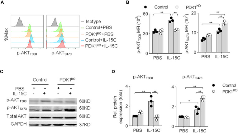
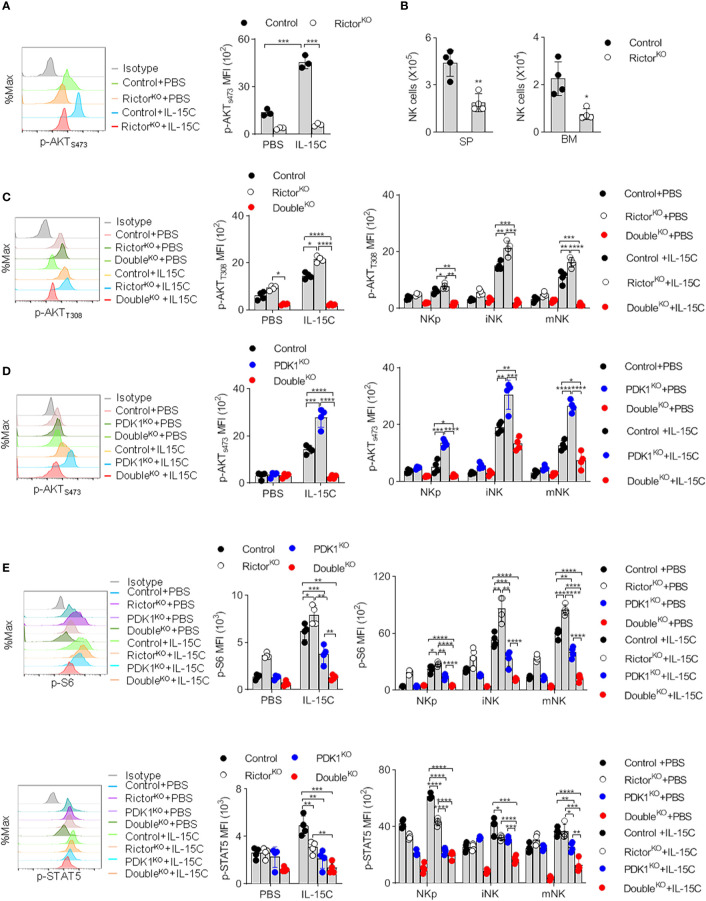
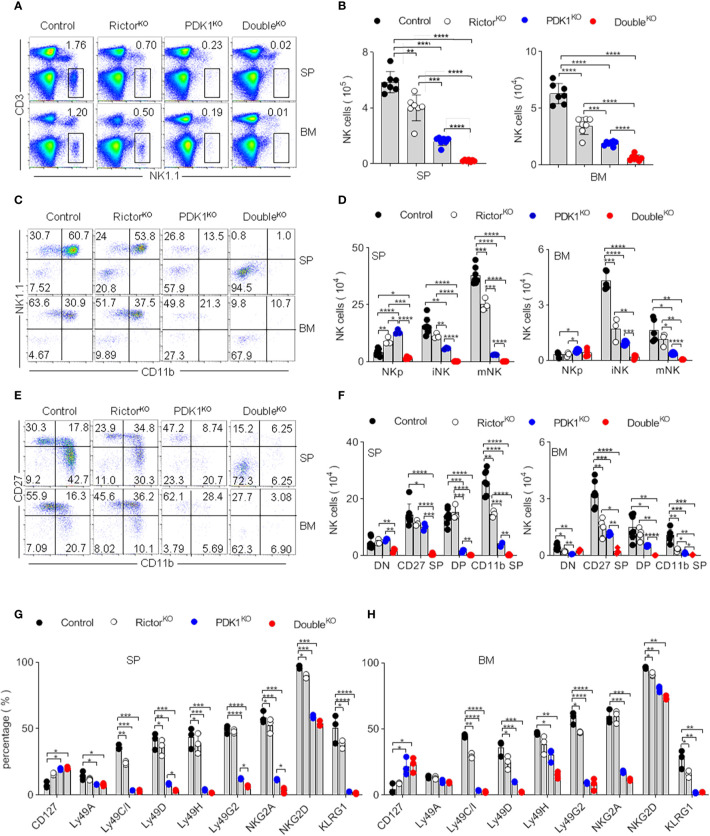
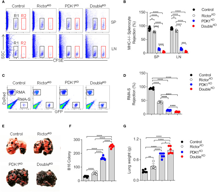
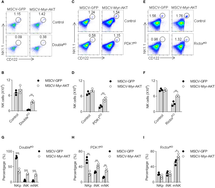
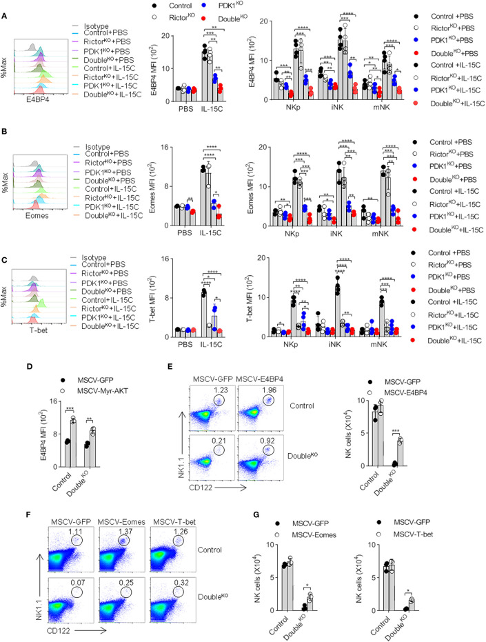
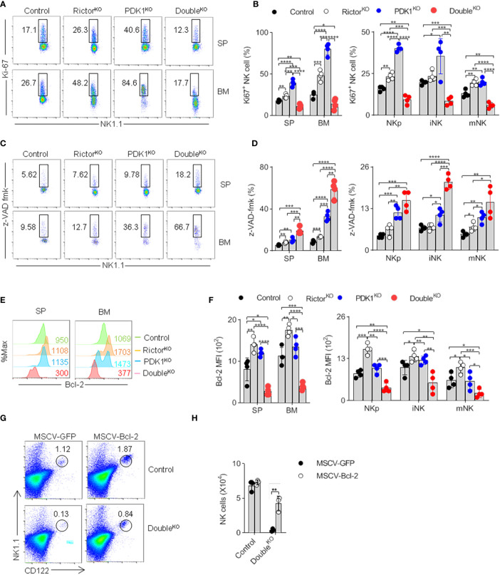
The role of PI3K-mTOR pathway in regulating NK cell development has been widely reported. However, it remains unclear whether NK cell development depends on the protein kinase B (PKB), which links PI3K and mTOR, perhaps due to the potential redundancy of PKB. PKB has two phosphorylation sites, threonine 308 (T308) and serine 473 (S473), which can be phosphorylated by phosphoinositide-dependent protein kinase-1 (PDK1) and mTORC2, respectively. In this study, we established a mouse model in which PKB was inactivated through the deletion of PDK1 and Rictor, a key component of mTORC2, respectively. We found that the single deletion of PDK1 or Rictor could lead to a significant defect in NK cell development, while combined deletion of PDK1 and Rictor severely hindered NK cell development at the early stage. Notably, ectopic expression of myristoylated PKB significantly rescued this defect. In terms of mechanism, in PDK1/Rictor-deficient NK cells, E4BP4, a transcription factor for NK cell development, was less expressed, and the exogenous supply of E4BP4 could alleviate the developmental defect of NK cell in these mice. Besides, overexpression of Bcl-2 also helped the survival of PDK1/Rictor-deficient NK cells, suggesting an anti-apoptotic role of PKB in NK cells. In summary, complete phosphorylation of PKB at T308 and S473 by PDK1 and mTORC2 is necessary for optimal NK cell development, and PKB regulates NK cell development by promoting E4BP4 expression and preventing cell apoptosis.

Keywords: development; mammalian target of rapamycin complex 2; natural killer (NK) cell; phosphoinositide-dependent protein kinase-1 (PDK1); protein kinase B (PKB); survival.


Conflict of interest statement

The authors declare that the research was conducted in the absence of any commercial or financial relationships that could be construed as a potential conflict of interest.

Figures

Figure 1
Figure 1
Deletion of PDK1 abolishes PKB T308 phosphorylation but enhances PKB S473 phosphorylation in NK cells. (A, B) The expression levels of AKT phosphorylated at T308 (p-AKTT308) and S473 (p-AKTS473) in Control or PDK1-deficient NK cells were determined by flow cytometry after IL-15C stimulation. Representative overlaid histograms (A), absolute MFI for p-AKTT308 and p-AKTS473 (B). (C, D) The expression levels of p-AKTT308 and p-AKTS473 in Control or PDK1-deficient NK cell were analyzed by Western blotting (C) and densitometry (D) after IL-15C stimulation. *p < 0.05, **p < 0.01.
Figure 2
Figure 2
Deletion of Rictor abolishes the enhanced phosphorylation of PKB S473 in PDK1-deficient NK cells. (A) The expression levels of p-AKTS473 in Rictor-deficient NK cell were detected by flow cytometry after IL-15C stimulation. (B) The number of NK cell in the spleen (SP) and bone marrow (BM) of Rictor-deficient mice was counted. (C) The expression levels of p-AKTT308 in Control, Rictor-deficient or PDK1/Rictor double deficient NK cells were detected by flow cytometry after IL-15C stimulation. (D) The expression levels of p-AKTS473 in Control, PDK1-deficient or PDK1/Rictor double deficient NK cells were determined by flow cytometry after IL-15C stimulation. (E) The expression levels of p-S6 and p-STAT5 in spleen NK cell were detected by flow cytometry after IL-15C stimulation in indicated mice. Representative overlaid histograms (left), absolute MFI (right).The data represent one of three independent experiments, n≥3 for each experiment, and values are expressed as the mean ± SD. *p < 0.05, **p < 0.01, ***p < 0.001, ****p < 0.0001.
Figure 3
Figure 3
mTORC2 deficiency aggravates the developmental defect of PDK1-deficient NK cells. (A, B) Representative flow cytometric plots (A) and the absolute number (B) of NK cell (CD3-NK1.1+) in the SP and BM of the indicated mice. (C, D) Representative flow cytometric plots (C) and the number (D) of NK cell subsets in the SP and BM of annotated mice. NKp (CD122+NK1.1-CD11b-), iNK (CD122+NK1.1+CD11b-), mNK (CD122+NK1.1+CD11b+). (E, F) Representative flow cytometric plots of CD27 and CD11b expression (E) and number of stage-specific NK cell (F) in the SP and BM of annotated mice. DN (CD27-CD11b-), CD27 SP (CD27+CD11b-), DP (CD27+CD11b+) and CD11b SP (CD27-CD11b+). (G, H) Statistical results of percentage in NK cell membrane receptor expression spectra in SP (G) and BM (H) of indicated mice. The data represent one of three independent experiments, n≥3 for each experiment, and values are expressed as the mean ± SD. *p < 0.05, **p < 0.01, ***p < 0.001, ****p < 0.0001.
Figure 4
Figure 4
NK cells fail to mediate in vivo surveillance in PDK1/Rictor-deficient mice. (A) Representative flow cytometry plot of CFSE+ cells obtained from the SP and Lymph nodes (LN) of the indicated recipient mice at 18 h after injection with an equal number of C57BL/6 and β2m-deficient splenocytes labeled with various concentrations of the cytosolic dye CFSE. R1, CFSE-low splenocytes from C57BL/6 mice; and R2, CFSE-high splenocytes from β2m-deficient mice. (B) The percentages of rejected β2m-deficient splenocytes from the SP and LN of the indicated recipient mice. (C) Representative flow cytometry plot of injected RMA-S cells in the peritoneal cavity at 18 h after intraperitoneal injection of the indicated mice with a mixture of NK cell-sensitive RMA-S cells expressing green fluorescent protein (GFP) together with NK cell-resistant RMA cells expressing the fluorescent protein DsRed. (D) The percentage of rejected RMA-S cells. (E–G) B16 metastasis assay, the indicated mice were injected intravenously with 2×10⁵ B16 cells. The mice were sacrificed 14 day later, and the lung weights and numbers of tumor nodules were counted. Each symbol represents an individual mouse. Data are shown as means ± SD and represent one of three independent experiments, n≥3 for each experiment. *p < 0.05, **p < 0.01, ***p < 0.001, ****p < 0.0001.
Figure 5
Figure 5
The ectopic expression of myristoylated PKB corrects the defect of NK cell development. (A–F) Double- (A, B), PDK1- (C, D) or Rictor-deficient (E, F) mice were treated with 5-FU for 4 days, and bone marrow cells were collected for spin-infection with MSCV retrovirus encoding control GFP, or Myr-AKT (activated-PKB). The infected BM cells were then transferred into recipient mice. After 8 weeks, CD122 vs NK1.1 expression on gated CD3 cells was analyzed by flow cytometry. The numbers in the outlined areas indicate the percent of CD3CD122+NK1.1+ cells (A, C, E). Numbers of CD3CD122+NK1.1+ cell are enumerated (B, D, F). (G–I) The developmental stages of NK cell was analysed after replenish Myr-AKT to HSC of Double-, PDK1- or Rictor-deficient mice (G–I). The data represent one of three independent experiments, n≥3 for each experiment, and values are expressed as the mean ± SD. *p < 0.05, **p < 0.01, ***p < 0.001.
Figure 6
Figure 6
PDK1 and mTORC2 coordinate to regulate the expression of E4BP4 by activating PKB. (A–C) The expressions of E4BP4 (A), Eomes (B), and T-bet (C) in total NK cells and NK cells at different stages in mice spleen were examined by flow cytometry. Representative overlaid histograms (left), the absolute MFI in total NK cell (middle) and NK cell subsets (left) from mice as annotated. (D) The expression level of E4BP4 in NK cell was examined after replenish Myr-AKT to HSC of DoubleKO mice. (E–G) The percentage and number of NK cell were analyzed after replenish E4BP4 (E), Eomes or T-bet (F, G) to HSC of DoubleKO mice. The data represent one of three independent experiments, n≥3 for each experiment, and values are expressed as the mean ± SD. *p < 0.05, **p < 0.01, ***p < 0.001, ****p < 0.0001.
Figure 7
Figure 7
Reduced PKB activation promotes NK cell apoptosis. (A, B) Representative flowcytometric plots (A) and the percentage (B) of total and stage-specific Ki67+ NK cells in the SP and BM of indicated mice. (C, D) Representative flow cytometric plots (C) and the proportion (D) of total and stage-specific caspase-positive NK cell in the SP and BM of indicated mice. (E, F) Representative overlaid histograms (E) and the absolute MFI (F) demonstrate the expression levels of Bcl-2 in total and stage-specific NK cells in the spleen and bone marrow of indicated-mice. (G, H) Representative flow cytometric results (G) and the statistics (H) indicate the effect of Bcl-2 on the proportion of NK cell after supplement Bcl-2 to the PDK1/Rictor-deficient mice. The data represent one of three independent experiments, n≥3 for each experiment, and values are expressed as the mean ± SD. *p < 0.05, **p < 0.01, ***p < 0.001, ****p < 0.0001.

Similar articles

Cited by

References

    1. Parham P, Guethlein LA. Genetics of Natural Killer Cells in Human Health, Disease,andSurvival. Annu Rev Immunol (2018) 36:519–48. 10.1146/annurev-immunol-042617-053149 - DOI - PubMed
    1. Vivier E, Tomasello E, Baratin M, Walzer T, Ugolini S. Functions of natural killer cells. Nat Immunol (2008) 9(5):503–10. 10.1038/ni1582 - DOI - PubMed
    1. Cooper MA, Bush JE, Fehniger TA, VanDeusen JB, Waite RE, Liu Y, et al. In vivo evidence for a dependence on interleukin 15 for survival of natural killer cells. Blood (2002) 100(10):3633–8. 10.1182/blood-2001-12-0293 - DOI - PubMed
    1. Kennedy MK, Glaccum M, Brown SN, Butz EA, Viney JL, Embers M, et al. Reversible defects in natural killer and memory CD8 T cell lineages in interleukin 15-deficient mice. J Exp Med (2000) 191(5):771–80. 10.1084/jem.191.5 - DOI - PMC - PubMed
    1. Ali AK, Nandagopal N, Lee SH. IL-15-PI3K-AKT-mTOR: A Critical Pathway in the Life Journey of Natural Killer Cells. Front Immunol (2015) 6:355:355. 10.3389/fimmu.2015.00355 - DOI - PMC - PubMed

Publication types

MeSH terms

Substances

LinkOut - more resources