Review
. 2021 Jan 28:11:625833.
doi: 10.3389/fimmu.2020.625833. eCollection 2020.

Signaling Through Nucleic Acid Sensors and Their Roles in Inflammatory Diseases

Affiliations
Review

Signaling Through Nucleic Acid Sensors and Their Roles in Inflammatory Diseases

Haruna Okude et al. Front Immunol. .

Abstract

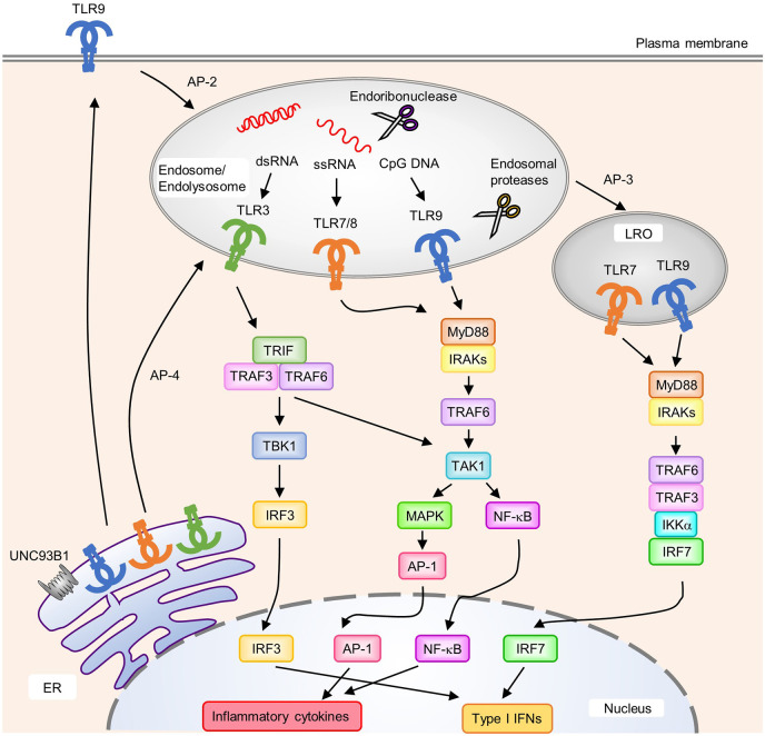
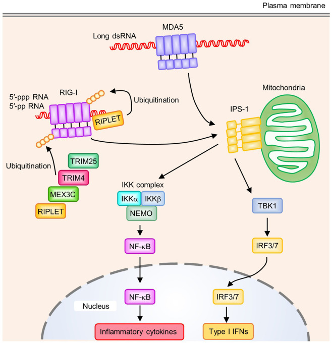
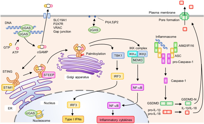
Recognition of pathogen-derived nucleic acids by pattern-recognition receptors (PRRs) is essential for eliciting antiviral immune responses by inducing the production of type I interferons (IFNs) and proinflammatory cytokines. Such responses are a prerequisite for mounting innate and pathogen-specific adaptive immune responses. However, host cells also use nucleic acids as carriers of genetic information, and the aberrant recognition of self-nucleic acids by PRRs is associated with the onset of autoimmune or autoinflammatory diseases. In this review, we describe the mechanisms of nucleic acid sensing by PRRs, including Toll-like receptors, RIG-I-like receptors, and DNA sensor molecules, and their signaling pathways as well as the disorders caused by uncontrolled or unnecessary activation of these PRRs.

Keywords: RIG-I-like receptor; Toll-like receptor; autoimmune disease; autoinflammatory disease; cGAMP synthase; nucleic acid sensing.


Conflict of interest statement

The authors declare that the research was conducted in the absence of any commercial or financial relationships that could be construed as a potential conflict of interest.

Figures

Figure 1
Figure 1
Localization, intracellular trafficking, and signaling pathways of nucleic acid-sensing Toll-like receptors (TLRs). TLR3, TLR7, TLR8, and TLR9 are synthesized in the endoplasmic reticulum (ER) and transported to endosomes via UNC93B1. Each TLR is transported to its destination [endosomes, endolysosomes, and lysosome-related organelles (LRO)] by individual mechanisms. TLR9 requires the AP-2 complex to translocate from the cell surface to endosomes, whereas TLR7 interacts with the AP-4 complex for direct trafficking to endosomes. Several endosomal proteases and endoribonucleases in endosomes/endolysosomes process TLRs and nucleic acids, respectively. Upon the recognition of cognate ligands, TLR7, TLR8, and TLR9 recruit MyD88 to activate downstream signaling pathways. MyD88 recruits IRAKs and TRAF6, which subsequently activate TAK1. Activated TAK1 leads to the activation of AP-1 through MAPK to initiate the transcription of proinflammatory cytokines. NF-κB is also activated by TAK1 and induces the production of proinflammatory cytokines. In pDCs, TLR7 and TLR9 in LRO induce the activation of IRF7 by forming a complex with TRAF6, TRAF3, IKKα; and IRF7, which results in the expression of type I IFNs. An AP-3 complex is required for the localization of TLR7 and TLR9 to LRO. TLR3 recruits TRIF to initiate downstream signaling pathway. TRIF recruits TRAF3 and TRAF6 to activate TBK1 and TAK1. Activated TBK1 induces type I IFNs through IRF3, and TAK1 induces proinflammatory cytokine production through NF-κB and AP-1.
Figure 2
Figure 2
Signaling pathways of RIG-I-like receptors (RLRs). RIG-I recognizes the 5′-tri- or 5′-di-phosphate end of RNA. A blunt-end at the triphosphate end and an unmethylated 5′-terminal nucleotide at the 2′-O position are also required to activate RIG-I. MDA5 binds to long dsRNA, which allows the oligomerization of MDA5 by forming a helical filament-like structure. Polyubiquitination on RIG-I CTD by RIPLET has a critical role in RIG-I activation. In addition to RIPLET, other E3 ubiquitin ligases such as TRIM25, TRIM4, and MEX3C act as positive regulators by mediating K63-linked polyubiquitination on RIG-I CARDs. Oligomerized CARDs of RIG-I or MDA5 interact with IPS-1 on mitochondria, which activates downstream signaling pathways. IPS-1 induces activation of the TBK1 and IKK complex (IKKα, IKKβ, and NEMO), which activates the transcription factors IRF3/7 and NF-κB, respectively. These transcription factors induce the production of type I IFNs and proinflammatory cytokines.
Figure 3
Figure 3
Activation of the cyclic GMP-AMP (cGAMP) synthase (cGAS)-stimulator of IFN genes (STING) signaling pathway and AIM2/IFI16 inflammasome. cGAS recognizes DNA in the cytosol and subsequently synthesizes the second messenger cGAMP from GTP and ATP. In the steady state, STIM1 interacts with STING to retain it on the endoplasmic reticulum (ER) membrane. Binding of cGAMP to STING induces the translocation of STING from the ER to the Golgi apparatus. Upon activation, enhanced interactions between STEEP and STING promote the trafficking of STING from the ER to the Golgi apparatus where it undergoes post-translational modifications such as palmitoylation. Activated STING on the Golgi apparatus recruits and activates TBK1 and the IKK complex (IKKα, IKKβ, and NEMO), which induce the production of type I IFNs and proinflammatory cytokines by IRF3 and NF-κB, respectively. cGAMP activates surrounding cells by being transferred to the extracellular space via SLC19A1, P2X7R, VRAC, and gap junctions. cGAS is associated with PI(4,5)P2 on the plasma membrane and is trafficked away from the nucleus to prevent the aberrant activation of cGAS by self-derived DNA. cGAS is also localized in the nucleus where its activity is inhibited by interactions with the nucleosome. Cytosolic DNA is recognized by ALRs, leading to the formation of an inflammasome composed of AIM2 or IFI16, ASC, and pro-Caspase-1. Within the inflammasome, Caspase-1 is activated by proteolytic cleavage from pro-Caspase-1 to Caspase-1. Activated Caspase-1 cleaves GSDMD, pro-IL-1β; and pro-IL-18. The N-terminus of GSDMD (GSDMD-N) forms a pore on the plasma membrane and induces cell death accompanied by the release of biologically active IL-1β and IL-18.

Similar articles

Cited by

References

    1. Brubaker SW, Bonham KS, Zanoni I, Kagan JC. Innate immune pattern recognition: a cell biological perspective. Annu Rev Immunol (2015) 33:257–90. 10.1146/annurev-immunol-032414-112240 - DOI - PMC - PubMed
    1. Kawasaki T, Kawai T, Akira S. Recognition of nucleic acids by pattern-recognition receptors and its relevance in autoimmunity. Immunol Rev (2011) 243(1):61–73. 10.1111/j.1600-065X.2011.01048.x - DOI - PMC - PubMed
    1. Alexopoulou L, Holt AC, Medzhitov R, Flavell RA. Recognition of double-stranded RNA and activation of NF-kappaB by Toll-like receptor 3. Nature (2001) 413(6857):732–8. 10.1038/35099560 - DOI - PubMed
    1. Hemmi H, Takeuchi O, Kawai T, Kaisho T, Sato S, Sanjo H, et al. A Toll-like receptor recognizes bacterial DNA. Nature (2000) 408(6813):740–5. 10.1038/35047123 - DOI - PubMed
    1. Heil F, Hemmi H, Hochrein H, Ampenberger F, Kirschning C, Akira S, et al. Species-specific recognition of single-stranded RNA via toll-like receptor 7 and 8. Science (2004) 303(5663):1526–9. 10.1126/science.1093620 - DOI - PubMed

LinkOut - more resources