. 2021 Feb 9:12:589518.
doi: 10.3389/fpls.2021.589518. eCollection 2021.

At FUT4 and At FUT6 Are Arabinofuranose-Specific Fucosyltransferases

Affiliations

At FUT4 and At FUT6 Are Arabinofuranose-Specific Fucosyltransferases

Maria J Soto et al. Front Plant Sci. .

Abstract

The bulk of plant biomass is comprised of plant cell walls, which are complex polymeric networks, composed of diverse polysaccharides, proteins, polyphenolics, and hydroxyproline-rich glycoproteins (HRGPs). Glycosyltransferases (GTs) work together to synthesize the saccharide components of the plant cell wall. The Arabidopsis thaliana fucosyltransferases (FUTs), AtFUT4, and AtFUT6, are members of the plant-specific GT family 37 (GT37). AtFUT4 and AtFUT6 transfer fucose (Fuc) onto arabinose (Ara) residues of arabinogalactan (AG) proteins (AGPs) and have been postulated to be non-redundant AGP-specific FUTs. AtFUT4 and AtFUT6 were recombinantly expressed in mammalian HEK293 cells and purified for biochemical analysis. We report an updated understanding on the specificities of AtFUT4 and AtFUT6 that are involved in the synthesis of wall localized AGPs. Our findings suggest that they are selective enzymes that can utilize various arabinogalactan (AG)-like and non-AG-like oligosaccharide acceptors, and only require a free, terminal arabinofuranose. We also report with GUS promoter-reporter gene studies that AtFUT4 and AtFUT6 gene expression is sub-localized in different parts of developing A. thaliana roots.

Keywords: AtFUT1; AtFUT4; AtFUT6; Fucosyltransferase; GT37; arabinogalactan protein; hydroxyproline-rich glycoprotein; plant cell wall.


Conflict of interest statement

The authors declare that the research was conducted in the absence of any commercial or financial relationships that could be construed as a potential conflict of interest.

Figures

Figure 1
Figure 1
Scheme showing the structures of xyloglucan (XyG) and arabinogalactan (AG) proteins (AGPs) from Arabidopsis thaliana with the predicted activities of AtFUT1 and AtFUT4/AtFUT6. (A) Representation of A. thaliana AGPs, and the putative activities of AtFUT4 and AtFUT6. Hyp, hydroxyproline. (B) Fucosylated XyG oligosaccharides derived from A. thaliana XyG (Peña et al., 2012; Tuomivaara et al., 2015). (C) Fucosylated AGP side chains derived from A. thaliana AGPs (Tryfona et al., 2012, 2014). (D) Symbol legend.
Figure 2
Figure 2
Structures of oligosaccharide acceptor substrates used in this study. (A) Synthetic arabinogalactan oligosaccharides numbered according to Ruprecht et al. (2020). (B) α-(1,5)-linked arabinan oligosaccharides. (C) Xyloglucan XXLG oligosaccharide. (D) β-(1,3)-linked galactobiose and β-(1,6)-linked galactobiose. (E) Symbol legend.
Figure 3
Figure 3
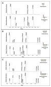
Matrix-assisted laser desorption/ionization-time of flight mass spectrometry (MALDI-TOF MS) data for GFP-AtFUT1, GFP-AtFUT4, and GFP-AtFUT6 reacted with different acceptor substrates. (A) Acceptor 55, (B) Acceptor 65, (C) Acceptor 68, (D) Acceptor 69, (E) Acceptor 70, and (F) XG acceptors. Transfer of Fuc increases the mass of the acceptor by 146 Da, as indicated by annotating (M + H+) ions, (*) denotes (M + Na+) adducts.
Figure 4
Figure 4
Biochemical analysis of GFP-AtFUT1, GFP-AtFUT4, and GFP-AtFUT6. Enzymatic activity was measured based on the production of GDP using the GDP-Glo assay kit in the presence or absence (Hydrolysis) of acceptor substrates (Figure 2). Values are represented as the average of three technical replicates ±SD, and are reported in nmol/min/mg of protein.
Figure 5
Figure 5
Relative enzyme activity of GFP-AtFUT1, GFP-AtFUT4, and GFP-AtFUT6 using AGPs extracted from vegetative tissue of wild-type (WT), fut4, fut6, and fut4fut6 A. thaliana mutants as acceptor substrates. Values are represented as the average of three technical replicates ±SD, and are reported in nmol/min/mg of protein.
Figure 6
Figure 6
MALDI-TOF MS analysis of products generated by incubating GFP-AtFUT4, and GFP-AtFUT6 with GDP-Fuc and different linear arabinan or galactan acceptor substrates. (A) β-(1,3)-linked galactobiose, (B) β-(1,6)-linked galactobiose, (C) α-(1,5)-linked arabinobiose, (D) α-(1,5)-linked, arabinotriose, and (E) α-(1,5)-linked arabinotetraose. Transfer of Fuc increases the mass of the acceptor by 146 Da, as indicated by annotating (M + H+) ions, (*) denotes (M + Na+) adducts. Control assays contained the same components except for enzyme.
Figure 7
Figure 7
NMR analysis of the products formed when arabinotriose was incubated with GFP-AtFUT4 and GDP-Fuc. The scheme of the reaction is shown at the top of the figure. The labeled cross-peaks in the two-dimensional COSY spectrum of the control (A) correspond to the anomeric signals of the residues of the arabinotriose. After the reaction with GFP-AtFUT4, the COSY spectrum (B) contained two additional signals, which were identified as terminal Fuc and an Ara with Fuc attached at O2. The signals surrounded by squares in the NOESY spectrum of the GFP-AtFUT4 reaction (C) indicate the glycosidic linkages in the enzymatically-generated fucosylated oligosaccharide. For the complete list of assignments, see Supplementary Table S2.
Figure 8
Figure 8
Linkage analysis of the reaction products of arabinan oligosaccharides incubated with GFP-AtFUT4. (A) Unreacted arabinobiose and arabinobiose incubated with GFP-AtFUT4. (B) Unreacted arabinotriose and arabinotriose incubated with GFP-AtFUT4. (C) Unreacted arabinotetraose and arabinotetraose incubated with GFP-AtFUT4. The presence of the 1,2-Araf peak in the spectra of the reaction products indicates that Fuc is attached to the terminal non-reducing Ara in all the oligosaccharides. As the 1,3,5-Araf and the 1,2,5-Araf peaks appear in the unreacted and reacted spectra we do not ascribe these to transfer in those linkages.
Figure 9
Figure 9
Imaging and staining of 12-day-old AtFUT4::GUS and AtFUT6::GUS seedlings. Multiple (n > 15) independent AtFUT4::GUS and AtFUT6::GUS plant lines are generated, grown, and observed for each construct. Visualization and qualitative analysis of GUS staining patterns of roots for two representative lines are shown. Staining patterns for lateral roots of increasing lengths are shown in (A,B,D,E), while close-ups of tap roots are shown in (C,F). (A,B) AtFUT4::GUS-1, (C) AtFUT4::GUS-2, (D,E) AtFUT6::GUS-1, and (F) AtFUT6::GUS-2.

Similar articles

Cited by

References

    1. Atmodjo M. A., Hao Z., Mohnen D. (2013). Evolving views of pectin biosynthesis. Annu. Rev. Plant Biol. 64, 747–779. 10.1146/annurev-arplant-042811-105534, PMID: - DOI - PubMed
    1. Bartetzko M. P., Pfrengle F. (2019). Automated glycan assembly of plant oligosaccharides and their application in cell-wall biology. ChemBioChem 20, 877–885. 10.1002/cbic.201800641, PMID: - DOI - PubMed
    1. Bartetzko M. P., Schuhmacher F., Hahm H. S., Seeberger P. H., Pfrengle F. (2015). Automated glycan assembly of oligosaccharides related to arabinogalactan proteins. Org. Lett. 17, 4344–4347. 10.1021/acs.orglett.5b02185, PMID: - DOI - PubMed
    1. Bonin C. P., Freshour G., Hahn M. G., Vanzin G. F., Reiter W. -D., Hahn M. G. (2003). The GMD1 and GMD2 genes of Arabidopsis encode isoforms of GDP-D-mannose 4,6-dehydratase with cell type-specific expression patterns. Plant Physiol. 132, 883–892. 10.1104/pp.103.022368, PMID: - DOI - PMC - PubMed
    1. Bonin C. P., Potter I., Vanzin G. F., Reiter W. D. (1997). The MUR1 gene of Arabidopsis thaliana encodes an isoform of GDP-d-mannose-4,6-dehydratase, catalyzing the first step in the de novo synthesis of GDP-l-fucose. Proc. Natl. Acad. Sci. U. S. A. 94, 2085–2090. 10.1073/PNAS.94.5.2085, PMID: - DOI - PMC - PubMed