. 2021 Feb 3:11:627007.
doi: 10.3389/fgene.2020.627007. eCollection 2020.

Human Ubiquitin-Specific Peptidase 18 Is Regulated by microRNAs via the 3'Untranslated Region, A Sequence Duplicated in Long Intergenic Non-coding RNA Genes Residing in chr22q11.21

Affiliations

Human Ubiquitin-Specific Peptidase 18 Is Regulated by microRNAs via the 3'Untranslated Region, A Sequence Duplicated in Long Intergenic Non-coding RNA Genes Residing in chr22q11.21

Erminia Rubino et al. Front Genet. .

Abstract

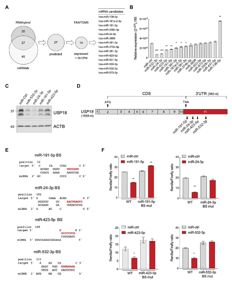
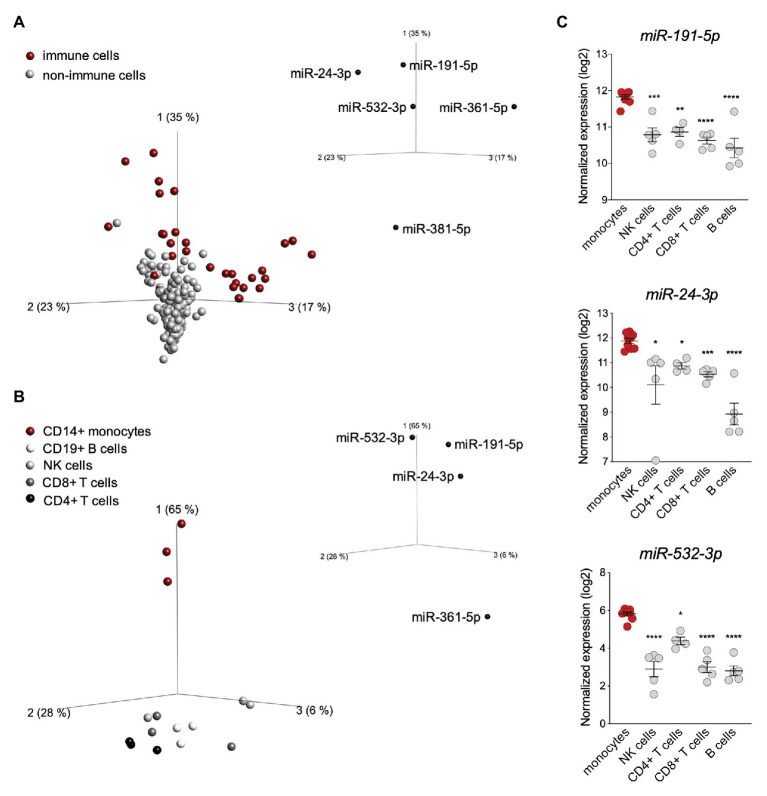
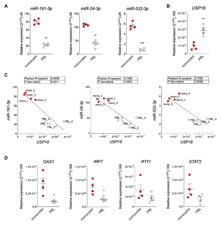
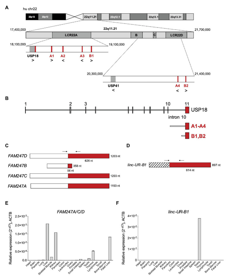
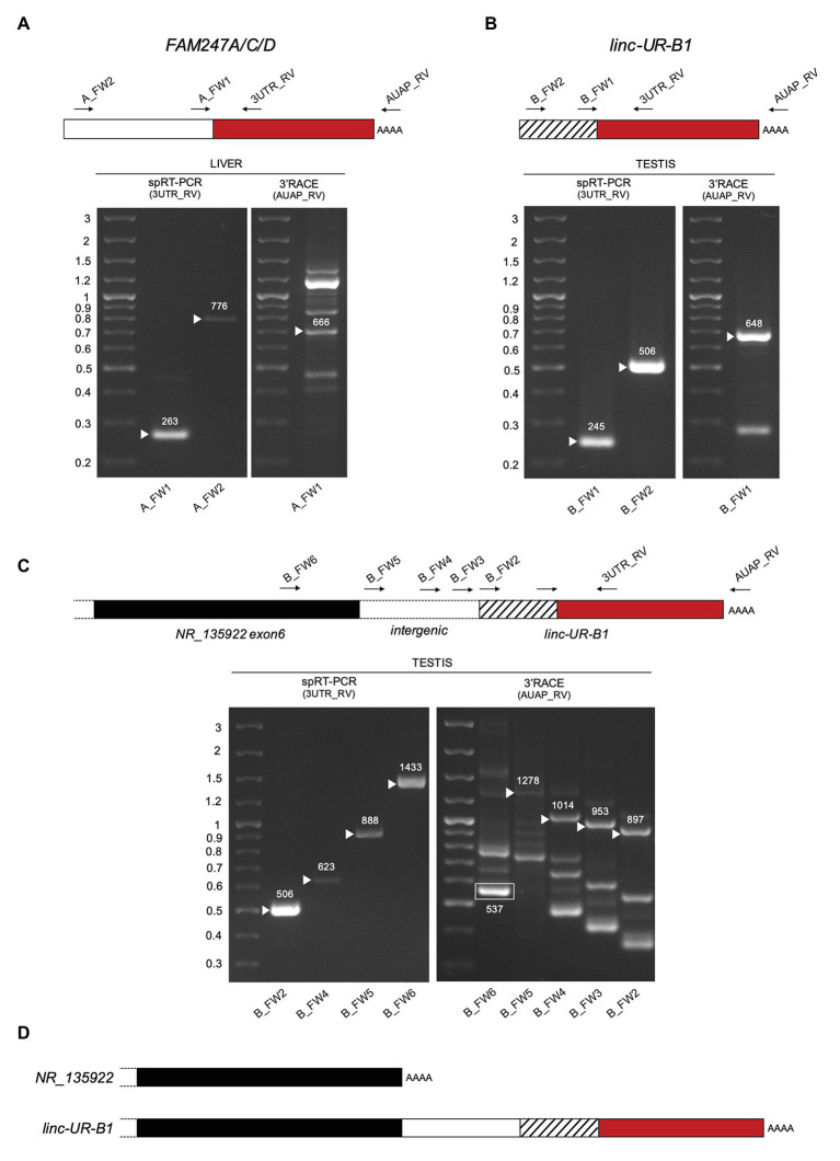
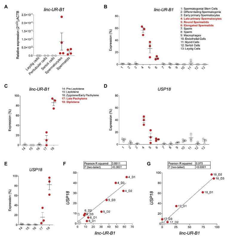
Ubiquitin-specific peptidase 18 (USP18) acts as gatekeeper of type I interferon (IFN) responses by binding to the IFN receptor subunit IFNAR2 and preventing activation of the downstream JAK/STAT pathway. In any given cell type, the level of USP18 is a key determinant of the output of IFN-stimulated transcripts. How the baseline level of USP18 is finely tuned in different cell types remains ill defined. Here, we identified microRNAs (miRNAs) that efficiently target USP18 through binding to the 3'untranslated region (3'UTR). Among these, three miRNAs are particularly enriched in circulating monocytes which exhibit low baseline USP18. Intriguingly, the USP18 3'UTR sequence is duplicated in human and chimpanzee genomes. In humans, four USP18 3'UTR copies were previously found to be embedded in long intergenic non-coding (linc) RNA genes residing in chr22q11.21 and known as FAM247A-D. Here, we further characterized their sequence and measured their expression profile in human tissues. Importantly, we describe an additional lincRNA bearing USP18 3'UTR (here linc-UR-B1) that is expressed only in testis. RNA-seq data analyses from testicular cell subsets revealed a positive correlation between linc-UR-B1 and USP18 expression in spermatocytes and spermatids. Overall, our findings uncover a set of miRNAs and lincRNAs, which may be part of a network evolved to fine-tune baseline USP18, particularly in cell types where IFN responsiveness needs to be tightly controlled.

Keywords: 22q11.2; 3'untranslated region, microRNAs; Testis; long intergenic non-coding RNA; type I interferon; ubiquitin-specific peptidase.


Conflict of interest statement

The authors declare that the research was conducted in the absence of any commercial or financial relationships that could be construed as a potential conflict of interest.

Figures

Figure 1
Figure 1
Identification of USP18-targeting miRNAs. (A) Prediction of USP18-targeting miRNAs using RNA hybrid algorithm and mirWalk software (TargetScan, Pita, miRDB, and RNA22). The common high-scoring miRNAs (27) were further selected for expression (counts per million mapped reads, CPM > 50) in 90 human cell types (FANTOM5 dataset). The selected candidates (14) are listed. (B) The 14 selected miRNAs and a miR-control (miR-ctrl) were expressed as mimics in HeLa S3 cells and USP18 mRNA was measured by qPCR (n = 4). Results (± SEM) shown as expression (2-ΔCt) relative to 18S, used as housekeeping gene. Raw-matched one-way ANOVA with Geisser-Greenhouse correction was performed (Dunnett’s multiple comparison – compared to miR-ctrl), adjusted values shown as stars *p < 0.05, **p < 0.01, ***p < 0.001, ****p < 0.0001. (C) The miR-ctrl and the indicated miRNAs were expressed as mimics in HeLa S3 cells and 48 h later endogenous USP18 was measured by western blot. Actin B (ACTB) as loading control. The data are representative of three experiments. (D) Schematic of the USP18 mRNA. Numbered boxes correspond to the exons. CDS, coding sequences. Arrowheads, the start (ATG) and stop (TAA) codons and the binding sites for the four indicated miRNAs. Note that exon 11 (in red) contains 43 coding nt, the stop codon, and the 3’untranslated region (UTR). The sequences of USP18 exon11 are provided as Supplementary Information. (E) Binding sites (BS) of miR-191-5p, miR-24-3p, miR-423-5p, and miR-532-3p in the USP18 3’UTR. Seed-matched sequences are in red. Pairing of miRNA-USP18 sequences was obtained with the RNAhybrid algorithm. (F) HeLa S3 cells were transfected with the indicated miRNAs and 24 h later with the psiCHECK2-USP18 3’UTR reporter plasmid, WT, or mutated in the miRNA binding site (BS mut). Each mutant plasmid contains a 5 nt mutation (nt 2–6 of the seed-matched sequence shown in E). Luciferase activity was measured 24 h after plasmid transfection (n = 4) and expressed as Renilla/Firefly ratio (± SEM).
Figure 2
Figure 2
miR-191-5p, miR-24-3p, and miR-532-3p are enriched in monocytes. (A) Principal component analysis (PCA) performed on 90 human cell types (FANTOM5 project dataset, three donors per cell population). The distribution is based on expression of the seven microRNAs (miRNAs) significantly targeting ubiquitin-specific peptidase 18 (USP18; see Figure 1B). Red and gray circles: immune and non-immune cells, respectively. The PCA plot shown captures 75% of the total variance within the selected data set (PCA1 35%, PCA2 23%, and PCA3 17%). value of (ANOVA FDR adjusted p-value) q < 0.05. The five USP18-targeting miRNA candidates significantly contributing to the data distribution are shown on the three PC axes of the plot. (B) PCA performed on circulating immune cell populations (FANTOM5 project data set). Analysis was restricted to the four USP18-targeting miRNAs enriched in immune cells. Color legend on the left. The PCA plot captures 97% of the total variance within the selected data set (PCA1 65%, PCA2 28%, and PCA3 6%). value of (ANOVA FDR adjusted p-value) q < 0.05. The four miRNAs significantly contributing to the data distribution are shown on the three principal component axes of the PCA plot. (C) Normalized expression (log2, ±SEM) of miR-532-3p, miR-191-5p, and miR-24-3p in circulating immune cell subsets. Data retrieved from (Allantaz et al., 2012), Cohort Roche, GSE28492. Ordinary one-way ANOVA (Dunnett’s multiple comparison – compared to monocytes) was performed, adjusted p-values shown as stars *p < 0.05, **p < 0.01, ***p < 0.001.
Figure 3
Figure 3
miR-191-5p, miR-24-3p, and miR-532-3p negatively correlate with USP18 in monocytes vs. PBL. (A,B) Expression of miR-191-5p, miR-24-3p, and miR-532-3p (A) and of USP18 (B) in ex vivo monocytes and monocyte-depleted peripheral blood mononuclear cells (PBMCs; here PBL; four donors). Results (± SEM) shown as expression (2-ΔCt) relative to U6 or 18S, used as housekeeping genes. Paired t-test values of p shown as stars **p < 0.01, ***p < 0.001. (C) Negative correlation between USP18 and miR-191-5p, miR-24-3p, and miR-532-3p in monocytes and PBL. Expression levels used to calculate the correlation are shown in (A,B). Pearson correlation coefficients and p-values (two-tailed) are shown above. (D) Expression of OAS1, IRF7, IFIT1, and STAT2 in monocytes and PBL (four donors). Results (± SEM) shown as expression (2-ΔCt) relative to 18S. Paired t test p-values are shown, when significant, as stars *p < 0.05.
Figure 4
Figure 4
Several USP18 exon 11 copies are embedded in expressed long intergenic non-coding RNA (lincRNA) genes. (A) Schematic of human chr22 (about 50 Mb) and below the chr22q11.21 region with four LCR22s. The bona fide USP18 gene resides at the boundary of LCR22A. The pseudogene USP41, located in LCR22B, contains most USP18 gene sequences, i.e., from exon 3 to exon 10, but lacks the 5’ and 3’ UTRs. The six copies of USP18 exon 11 are indicated in red. Genes and USP18 copies are shown with their genomic orientation (> for + strand, < for − strand). (B) Top, intron-exon organization of the human USP18 gene. Exons, gray boxes; introns, lines. Exon 11 is highlighted in red. Below are aligned the duplications found. Copies A1–4 contain most of USP18 intron 10 and the entire exon 11. Copies B1 and B2 contain a smaller sequence of intron 10 and exon 11. (C) Map of FAM247A-D transcripts containing USP18 exon 11 in red. The annotated exon upstream of exon 11 is represented as white box. Arrows indicate the primers (A_FW1 and A_RV1) designed across the junction and used to measure expression of FAM247A/C/D in (E). (D) Map of linc-UR-B1. The annotated exon upstream of exon 11 is represented as striped box. Arrows indicate the primers (B_FW1 and B_RV1) designed across the junction and used to measure expression of linc-UR-B1 in (F). (E) Expression of FAM247A/C/D in 20 human tissues (pool of donors for each tissue; Human Immune System MTC™ Panel, Human MTC™ Panel I, and Human MTC™ Panel II). Results shown as expression (2-ΔCt) relative to ACTB. SEM is shown for tissues present in two of the above panels. (F) Expression of linc-UR-B1 in the same samples used in (E).
Figure 5
Figure 5
Characterization of FAM247A/C/D and linc-UR-B1 molecules. (A) Analysis of FAM247 transcripts in liver. Right gel, 3’ of cDNA Ends (3’RACE). Complementary DNA (cDNA) was obtained by oligo-dT reverse transcription (RT) of polyA+ liver RNA. PCR was performed on this cDNA using the forward (A_FW1) and reverse (AUAP_RV) primers indicated on the schematic above. Several bands were obtained, including the expected 666 bp. Left gel, specific RT-PCR (spRT-PCR). cDNA was obtained from polyA+ liver RNA using a primer designed on the USP18 3’UTR (3UTR_GSP1). PCR was then performed using the forward primer (A_FW1 or A_FW2) and a reverse primer designed on the 3’UTR (3UTR_RV) and internal to 3UTR_GSP1. The length in nt is indicated above each amplified product. (B) Analysis of linc-UR-B1 transcripts in testis. The strategies described in (A) were used on testis poly A+ RNA. Primers are indicated on the schematic above. The length in nt is indicated above each amplified band. (C) Study of linc-UR-B1 transcript extending on the 5’ end. Three forward primers were designed on the annotated intergenic region (B_FW3-4-5). One primer (B_FW6) was designed on the sequence further upstream, corresponding to exon 6 of the annotated NR_135922 transcript. Right panel, 3’RACE on testis RNA. The 537 nt product obtained using the B_FW6 primer corresponds to the annotated NR_135922. Longer products indicated by arrowheads were obtained when using the three primers designed in the intergenic region. Left panel, spRT-PCR was performed on testis RNA using the indicated forward primers and the 3UTR_RV primer. PCR products of the expected lengths were obtained for all primers tested. Panels (A–C): the PCR products of the size expected from the annotations were sequenced. The additional 3’RACE PCR products likely result from the amplification of other transcripts containing sequences identical to the exon upstream of USP18 exon11 (Supplementary Table S1). (D) Schematic of the 3’ end of the two NR_135922 isoforms identified here. Top, the NR_135922 transcript terminating with exon 6, as in the annotation. Bottom, longer isoform of NR_135922 terminating with the USP18 3’UTR, here called linc-UR-B1. The sequences of FAM247A/C/D and linc-UR-B1 are provided as Supplementary Information.
Figure 6
Figure 6
linc-UR-B1 is expressed in meiotic and post-meiotic germ cells and positively correlates with USP18. (A) Measure of linc-UR-B1 levels by quantitative PCR (qPCR) in purified testicular cell populations (spermatocytes, spermatids, peritubular cells, and Leydig cells) and Sertoli cells. Results (± SEM) shown as expression (2-ΔCt) relative to ACTB. (B,D) Analysis of linc-UR-B1 (B) and USP18 (D) expression in testicular cell populations (clusters 1–13, three donors). (C,E) Analysis of linc-UR-B1 (C) and USP18 (E) expression in clusters of germ cells populations at different stages of prophase I (three donors). Note that clusters 14–18 are subclusters of clusters 3 and 4 studied in (B) and (D). (F,G) Positive correlation of linc-UR-B1 and USP18 expression in germ cell populations (F, clusters 1–8) and in germ cells at different stages of prophase I (G, clusters 14–18). All data were retrieved from the alignment of single cell RNA-seq reads provided by (Guo et al., 2018), filtered for unique reads, and expressed (± SEM) as percentage expression (normalized expression in one population vs. all populations analyzed). Cell populations expressing linc-UR-B1 are highlighted in red.

Similar articles

Cited by

References

    1. Allantaz F., Cheng D. T., Bergauer T., Ravindran P., Rossier M. F., Ebeling M., et al. . (2012). Expression profiling of human immune cell subsets identifies miRNA-mRNA regulatory relationships correlated with cell type specific expression. PLoS One 7:e29979. 10.1371/journal.pone.0029979, PMID: - DOI - PMC - PubMed
    1. Arimoto K. I., Lochte S., Stoner S. A., Burkart C., Zhang Y., Miyauchi S., et al. . (2017). STAT2 is an essential adaptor in USP18-mediated suppression of type I interferon signaling. Nat. Struct. Mol. Biol. 24, 279–289. 10.1038/nsmb.3378, PMID: - DOI - PMC - PubMed
    1. Babcock M., Pavlicek A., Spiteri E., Kashork C. D., Ioshikhes I., Shaffer L. G., et al. . (2003). Shuffling of genes within low-copy repeats on 22q11 (LCR22) by Alu-mediated recombination events during evolution. Genome Res. 13, 2519–2532. 10.1101/gr.1549503, PMID: - DOI - PMC - PubMed
    1. Babcock M., Yatsenko S., Hopkins J., Brenton M., Cao Q., de Jong P., et al. . (2007). Hominoid lineage specific amplification of low-copy repeats on 22q11.2 (LCR22s) associated with velo-cardio-facial/digeorge syndrome. Hum. Mol. Genet. 16, 2560–2571. 10.1093/hmg/ddm197, PMID: - DOI - PubMed
    1. Bartel D. P. (2004). MicroRNAs: genomics, biogenesis, mechanism, and function. Cell 116, 281–297. 10.1016/S0092-8674(04)00045-5, PMID: - DOI - PubMed

LinkOut - more resources