

Usual Presentation Has Odds: Unilateral Tibial Hemimelia in One of Dizygotic Twins
- PMID: 33633877
- PMCID: PMC7899282
- DOI: 10.7759/cureus.12834
Usual Presentation Has Odds: Unilateral Tibial Hemimelia in One of Dizygotic Twins
Abstract
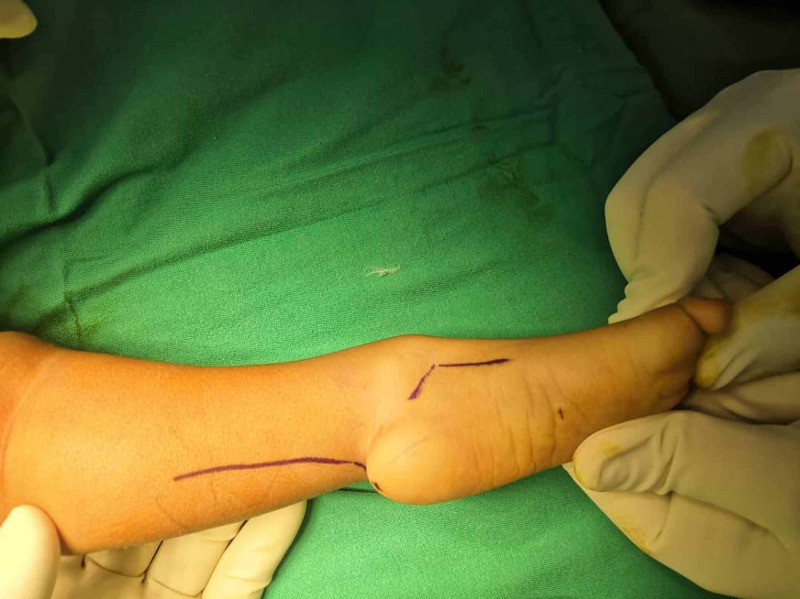
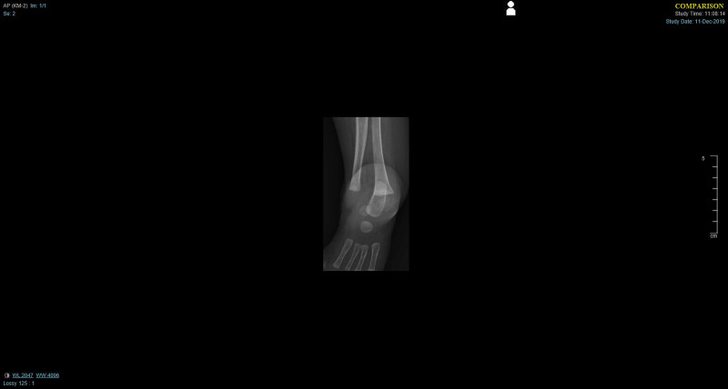
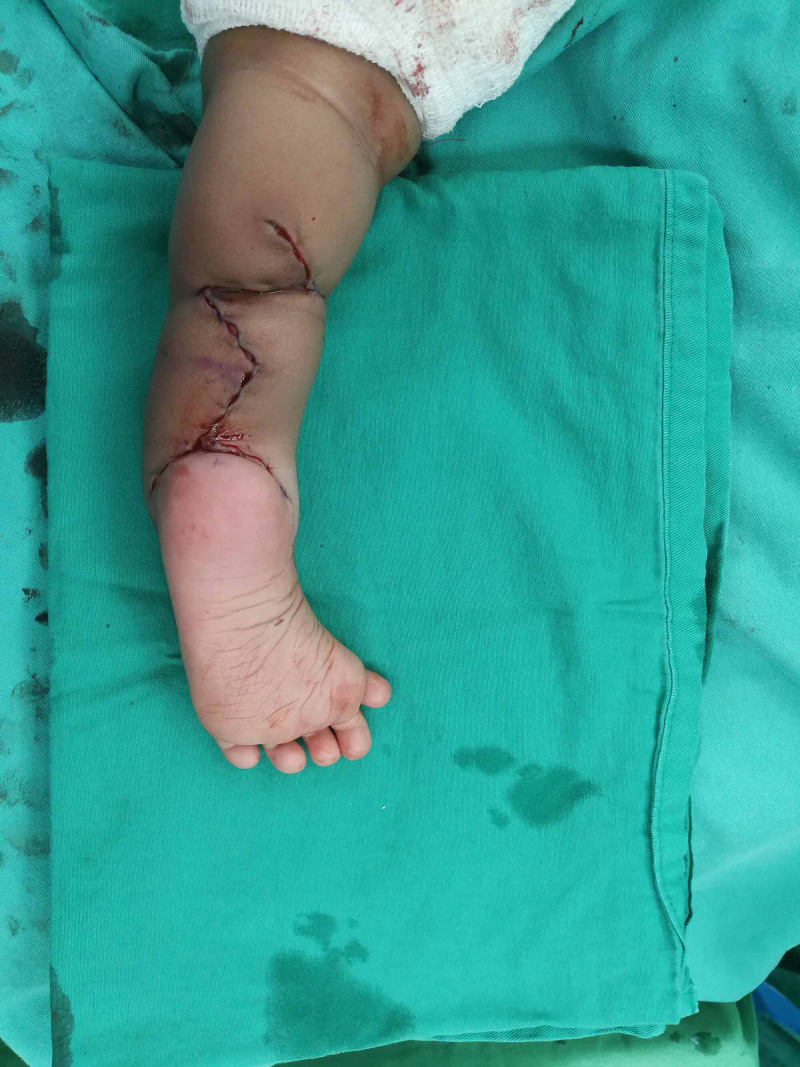
Tibial hemimelia is a relatively rare congenital tibial longitudinal deficiency (approximately 1 per 1 million live births), unilateral or bilateral, with a relatively intact fibula. Hemimelia results from a disruption of the lower limb developmental field during embryogenesis due to slow or even abort of chondrification process, which results in leg length discrepancy. Affected leg commonly appears short and deformed with knee, ankle, and foot involvement. It may present with a variety of associated anomalies. Surgical treatment varies according to the type and degree of deformity, and reconstructive interventions are still limited. Reported cases of tibial hemimelia are very infrequent, especially tibial hemimelia in twins. Usually, the cases were in single embryo or less frequently in one of the monozygotic twins, but no reported cases regarding tibial hemimelia in one of the dizygotic twins as this article reports.
Keywords: congenital tibial deficiency; hemimelia; tibial hemimelia.
Copyright © 2021, Al-Chalabi et al.
Conflict of interest statement
The authors have declared that no competing interests exist.
Figures
References
-
- Fibular hemimelia: more than just an absent bone. Fordham LA, Applegate KE, Wilkes DC, Chung CJ. Semin Musculoskelet Radiol. 1999;3:227–237. - PubMed
-
- A successful reconstructive case of tibia hemimelia in syndromic child. Matardiah NAM, Johari N, Hamid MA. Biomed J Sci Tech Res. 2019;19
-
- Tibial hemimelia in one of the identical twins. Leite JA, Lima LC, Sampaio ML. J Pediatr Orthop. 2010;30:742–745. - PubMed
-
- Systematic radiographic evaluation of tibial hemimelia with orthopedic implications. Kaplan-List K, Klionsky NB, Sanders JO, Katz ME. Pediatr Radiol. 2017;47:473–483. - PubMed
Publication types
LinkOut - more resources
Full Text Sources
Other Literature Sources