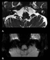
Primary Lymphoma of Internal Acoustic Meatus Mimicking Vestibular Schwannoma-A Rare Diagnostic Dilemma
- PMID: 33633923
- PMCID: PMC7902110
- DOI: 10.1055/s-0040-1722343
Primary Lymphoma of Internal Acoustic Meatus Mimicking Vestibular Schwannoma-A Rare Diagnostic Dilemma
Abstract
Background/Setting A subject presenting with a unilateral sensorineural hearing loss and with vertigo/imbalance and a lesion of internal acoustic meatus (IAM) most often represents a vestibular schwannoma. Several alternative pathologies involving the region, with clinical and neuroradiological similarities, could lead to an error in judgement and management. Rare tumors of the IAM pose unique diagnostic difficulty. A rare case that we present here had a typical history and imaging findings suggestive of vestibular schwannoma. A primary central nervous system (CNS) lymphoma was diagnosed in later stages of brain involvement warranting a retrospective analysis of the entity. Case Summary An 80-year-old male presented with unilateral sensorineural hearing loss, vertigo, and imbalance. On imaging, he was found to have a lesion in the left internal auditory meatus, reported as a vestibular schwannoma and operated upon. Subject's condition worsened with time and a repeat imaging was suggestive of a CNS lymphoma with lesions involving bilateral cerebellum and subcortical white matrix. Conclusion To conclude, primary CNS lymphoma presenting an isolated lesion in the IAM with no other parenchymal lesions at presentation is a rare incidence; to our knowledge this is the first case of such unique presentation.
Keywords: CNS lymphoma; imaging of internal acoustic meatus lesions; internal acoustic meatus; vestibular schwannoma.
The Author(s). This is an open access article published by Thieme under the terms of the Creative Commons Attribution-NonDerivative-NonCommercial License, permitting copying and reproduction so long as the original work is given appropriate credit. Contents may not be used for commercial purposes, or adapted, remixed, transformed or built upon. ( https://creativecommons.org/licenses/by-nc-nd/4.0/ ).
Conflict of interest statement
Conflict of Interest • The authors have no ethical conflicts to disclose. • The authors have no conflicts of interest to declare.
Figures


References
-
- Howitz M F, Johansen C, Tos M, Charabi S, Olsen J H. Incidence of vestibular schwannoma in Denmark, 1977-1995. Am J Otol. 2000;21(05):690–694. - PubMed
-
- Lin D, Hegarty J L, Fischbein N J, Jackler R K. The prevalence of “incidental” acoustic neuroma. Arch Otolaryngol Head Neck Surg. 2005;131(03):241–244. - PubMed
-
- Stangerup S E, Tos M, Thomsen J, Caye-Thomasen P.True incidence of vestibular schwannoma? Neurosurgery 201067051335–1340., discussion 1340 - PubMed
-
- Parker G D, Harnsberger H R. Clinical-radiologic issues in perineural tumor spread of malignant diseases of the extracranial head and neck. Radiographics. 1991;11(03):383–399. - PubMed
Publication types
LinkOut - more resources
Full Text Sources
Other Literature Sources

