

Prognostic analysis of pancreatic carcinoma with portal system invasion following curative resection
- PMID: 33633960
- PMCID: PMC7882355
- DOI: 10.21037/gs-20-495
Prognostic analysis of pancreatic carcinoma with portal system invasion following curative resection
Erratum in
-
Erratum to prognostic analysis of pancreatic carcinoma with portal system invasion following curative resection.Gland Surg. 2022 Sep;11(9):1586-1587. doi: 10.21037/gs-2022-03. Gland Surg. 2022. PMID: 36221272 Free PMC article.
Abstract
Background: To analyze the related factors affecting the prognosis of pancreatic carcinoma with portal system invasion.
Methods: We retrospectively analyzed the clinical data of 118 patients with portal venous system invasion in Beijing Chaoyang Hospital between January 2011 and December 2018. Only patients with borderline resectable pancreatic cancer were included in this study. Borderline pancreatic cancer was defined according to NCCN (The National Comprehensive Cancer Network) guidelines. All patients underwent surgical treatment combined with vascular resection and reconstruction. The prognosis was evaluated according to the follow-up results, and the related risk factors for prognosis were analyzed. The survival curve was drawn by Kaplan-Meier method, and the survival rate was compared by log-rank test. Multivariate Cox regression was used to analyze the prognostic factors.
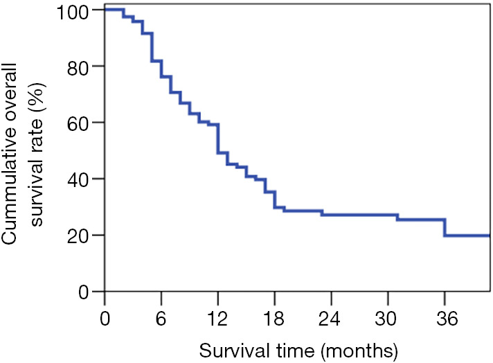
Results: In our research, all of 126 patients were successfully completed the operations. Complications occurred in 29.7% of patients and perioperative death in 4.0%. A total of 118 patients were followed up and the followed-up rate was 97.5% (118/121). The overall 1-year, 2-year and 3-year survival rates were 49.2%, 27.1% and 19.8%, And the median survival time was 20 months. Multivariate analysis showed that preoperative CA19-9 (RR 1.449, 95% CI: 1.053-1.994), N status (RR 2.533, 95% CI: 1.337-4.798), degree of tumor differentiation (RR 1.592, 95% CI: 1.064-2.381) and venous invasion depth (RR 2.03, 95% CI: 1.504-2.758) were independent risk factors for the prognosis.
Conclusions: The long-term prognosis of pancreatic carcinoma patients with portal system invasion is poor. The venous invasion depth is an independent risk factor for the prognosis of pancreatic carcinoma with portal system invasion, the deeper of venous invasion, the worse the prognosis, and poorly differentiated tumors have the worst prognosis. Other independent risk factors included N status and the preoperative CA19-9. Those may help with patients' selection for different treatment protocols.
Keywords: Pancreatic carcinoma; prognosis; risk factors; surgical procedures; vascular invasion.
2021 Gland Surgery. All rights reserved.
Conflict of interest statement
Conflicts of Interest: All authors have completed the ICMJE uniform disclosure form (available at http://dx.doi.org/10.21037/gs-20-495). The authors have no conflicts of interest to declare.
Figures


Comment in
-
Letter to the editor: the nonnegligible effect of neoadjuvant therapy for patients with borderline resectable pancreatic ductal adenocarcinoma.Gland Surg. 2021 Jul;10(7):2340-2342. doi: 10.21037/gs-21-379. Gland Surg. 2021. PMID: 34422605 Free PMC article. No abstract available.
References
LinkOut - more resources
Full Text Sources
Other Literature Sources