

Atypical enhanced computed tomography signs of pancreatic cancer and its differential diagnosis from autoimmune pancreatitis
- PMID: 33633991
- PMCID: PMC7882330
- DOI: 10.21037/gs-20-821
Atypical enhanced computed tomography signs of pancreatic cancer and its differential diagnosis from autoimmune pancreatitis
Abstract
Background: To analyze the atypical enhanced computed tomography (CT) signs of pancreatic cancer (PC) and compare them with those of autoimmune pancreatitis (AIP) to explore the differential diagnosis value of CT.
Methods: The clinical data of 36 AIP (AIP group) and 38 PC patients (PC group), who were admitted to our hospital from January 2013 to June 2020 and confirmed by surgical biopsy or hormone therapy, were retrospectively analyzed. Participants in both groups were examined by CT, the imaging signs of the 2 groups were analyzed, and the results of CT examination were compared.
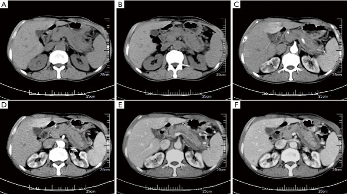
Results: In the PC group, the density of the lesions on the CT scan was mostly reduced, the pancreas was not swollen, and the kidneys were not involved. The bile duct wall was thickened with a sausage-like appearance, enveloped edges were rare, blood vessels were invaded, lymph nodes were enlarged, and the pancreatic duct was truncated. The findings of the AIP group were the opposite. The difference in the proportion of participants with the above-mentioned CT features between the 2 groups was statistically significant (P<0.05). The shape of the lesions in the AIP group was mainly elongated, of uneven density, and the density of enhanced scanning was medium to high. The predominant shape of the lesions in PC participants was spherical, and the density was uniform. The enhanced scan was mainly low-density. The difference in shape and density between the 2 groups was also statistically significant (P<0.05). The CT values of the plain scan, intravenous phase, and delayed phase in the AIP group were significantly higher than those in the PC group (P<0.05).
Conclusions: The imaging signs of AIP and PC overlap. Examination with CT is of great value in the differential diagnosis between AIP and PC. Familiarity with and mastery of the CT signs of AIP and PC can help to improve the accuracy of clinical diagnosis and provide a reliable basis for patients' follow-up treatment.
Keywords: Autoimmune pancreatitis (AIP); differential diagnosis; imaging signs; pancreatic cancer (PC).
2021 Gland Surgery. All rights reserved.
Conflict of interest statement
Conflicts of Interest: All authors have completed the ICMJE uniform disclosure form (available at http://dx.doi.org/10.21037/gs-20-821). The authors have no conflicts of interest to declare.
Figures
Similar articles
-
Focal autoimmune pancreatitis: radiological characteristics help to distinguish from pancreatic cancer.World J Gastroenterol. 2013 Jun 21;19(23):3634-41. doi: 10.3748/wjg.v19.i23.3634. World J Gastroenterol. 2013. PMID: 23801866 Free PMC article.
-
Differentiation of focal-type autoimmune pancreatitis from pancreatic carcinoma: assessment by multiphase contrast-enhanced CT.Eur Radiol. 2015 May;25(5):1366-74. doi: 10.1007/s00330-014-3512-3. Epub 2014 Nov 30. Eur Radiol. 2015. PMID: 25433412
-
18F- FDG PET/CT helps differentiate autoimmune pancreatitis from pancreatic cancer.BMC Cancer. 2017 Oct 23;17(1):695. doi: 10.1186/s12885-017-3665-y. BMC Cancer. 2017. PMID: 29061130 Free PMC article.
-
Imaging diagnosis of autoimmune pancreatitis using endoscopic ultrasonography.J Med Ultrason (2001). 2021 Oct;48(4):543-553. doi: 10.1007/s10396-021-01143-w. Epub 2021 Oct 20. J Med Ultrason (2001). 2021. PMID: 34669071 Review.
-
[Autoimmune pancreatitis: An update].Radiologe. 2016 Apr;56(4):363-70. doi: 10.1007/s00117-016-0096-8. Radiologe. 2016. PMID: 27025383 Review. German.
Cited by
-
Differentiation of autoimmune pancreatitis from pancreatic adenocarcinoma using CT characteristics: a systematic review and meta-analysis.Eur Radiol. 2023 Dec;33(12):9010-9021. doi: 10.1007/s00330-023-09959-5. Epub 2023 Jul 19. Eur Radiol. 2023. PMID: 37466708
-
Enhancing preoperative diagnosis of pancreatic ductal adenocarcinoma and mass-forming chronic pancreatitis: a study on normalized conventional MR imaging parameters.Abdom Radiol (NY). 2024 Nov 2. doi: 10.1007/s00261-024-04652-7. Online ahead of print. Abdom Radiol (NY). 2024. PMID: 39488674
-
Diagnostic Differentiation between Pancreatitis and Pancreatic Cancer: A Scoping Review.Diagnostics (Basel). 2024 Jan 29;14(3):290. doi: 10.3390/diagnostics14030290. Diagnostics (Basel). 2024. PMID: 38337806 Free PMC article.
References
LinkOut - more resources
Full Text Sources
Other Literature Sources
Miscellaneous