

Two cases of rare thyroid malignancy-case report
- PMID: 33633996
- PMCID: PMC7882315
- DOI: 10.21037/gs-20-601
Two cases of rare thyroid malignancy-case report
Abstract
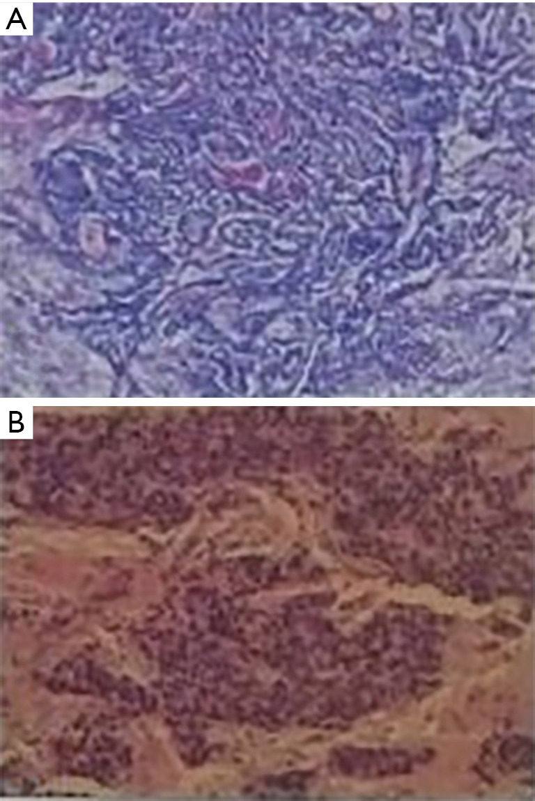
We report 2 cases of rare thyroid malignancy: angiosarcoma and myoepithelial carcinoma (MC). Thyroid angiosarcomas (TAS) is extremely rare and comprises less than 1% of primary thyroid cancer worldwide. MC usually presents as a slow-growing painless mass arising in the salivary glands. It has not been reported in the thyroid gland. The first case describes a 59-year-old patient who was admitted to hospital with the discovery of thyroid nodule for 1 month. The tumor thrombus was found in the left internal jugular vein and superior thyroid artery during the operation. Diagnosis of angiosarcoma of the thyroid was based on positive endothelial markers such as thrombomodulin and CD31 after total thyroidectomy. The left internal jugular vein, left recurrent laryngeal nerve and anterior cervical banding muscle were invaded by thyroid tumor. No lymph node metastasis was observed. The patient died after 4 years. The second case describes a 55-year-old woman who presented with the discovery of thyroid nodule for 1 month. Right thyroid lobectomy and right neck lymph node functional dissection were carried out. The results from postoperative pathology revealed that papillary carcinoma in right lobe of thyroid and MC next to thyroid were found. Besides, the metastasis of MC was observed at right II-IV level and right VI level. Five years later, the patient was re-admitted to hospital, primarily due to the discovery of anterior cervical tumor for one year. Then, she underwent left thyroid lobectomy and right tumor resection. Postoperative routine pathology showed recurrent MC in the right thyroid. After surgery and radiotherapy, the patient was followed up for 2 years. Angiosarcoma and myoepithelioma should be kept in mind in diagnosis of thyroid malignant tumor.
Keywords: Thyroid gland; angiosarcoma; case report; myoepithelial carcinoma (MC); thyroidectomy.
2021 Gland Surgery. All rights reserved.
Conflict of interest statement
Conflicts of Interest: All authors have completed the ICMJE uniform disclosure form (available at http://dx.doi.org/10.21037/gs-20-601). The authors have no conflicts of interest to declare.
Figures


Similar articles
-
Robot-assisted Sistrunk's operation, total thyroidectomy, and neck dissection via a transaxillary and retroauricular (TARA) approach in papillary carcinoma arising in thyroglossal duct cyst and thyroid gland.Ann Surg Oncol. 2012 Dec;19(13):4259-61. doi: 10.1245/s10434-012-2674-y. Epub 2012 Oct 16. Ann Surg Oncol. 2012. PMID: 23070784
-
Robotic total thyroidectomy with modified radical neck dissection via unilateral retroauricular approach.Ann Surg Oncol. 2014 Nov;21(12):3872-5. doi: 10.1245/s10434-014-3896-y. Epub 2014 Sep 17. Ann Surg Oncol. 2014. PMID: 25227305
-
A Rare Complication of the Thyroid Malignancies: Jugular Vein Invasion.Pol J Radiol. 2015 Jul 17;80:360-3. doi: 10.12659/PJR.894057. eCollection 2015. Pol J Radiol. 2015. PMID: 26236418 Free PMC article.
-
Breast cancer metastases to the thyroid gland - an uncommon sentinel for diffuse metastatic disease: a case report and review of the literature.J Med Case Rep. 2017 Sep 22;11(1):269. doi: 10.1186/s13256-017-1441-x. J Med Case Rep. 2017. PMID: 28934992 Free PMC article. Review.
-
Sclerosing mucoepidermoid carcinoma with eosinophilia of the thyroid: report of two patients, one with distant metastasis, and review of the literature.Hum Pathol. 1997 Sep;28(9):1091-6. doi: 10.1016/s0046-8177(97)90064-2. Hum Pathol. 1997. PMID: 9308735 Review.
Cited by
-
Thyroid angiosarcoma: a rare malignancy - case report and systematic review of the literature.Thyroid Res. 2025 Aug 1;18(1):38. doi: 10.1186/s13044-025-00252-9. Thyroid Res. 2025. PMID: 40745320 Free PMC article. Review.
References
-
- Li H, Li HX, Zhu Y, et al. Clinicopathologic study of primary angiosarcoma of thyroid. Zhonghua Bing Li Xue Za Zhi 2016;45:364-7. - PubMed
-
- Shen Z, Mao W, Zhang W. Retrospective analysis for 75 cases of angiosarcoma. Chin J Diffic and Compl Cas 2007;6:469-71.
-
- Surov A, Gottschling S, Wienke A, et al. Primary Thyroid Sarcoma: A Systematic Review. Anticancer Res 2015;35:5185-91. - PubMed
Publication types
LinkOut - more resources
Full Text Sources
Other Literature Sources