Review
. 2021 Jan;10(1):444-459.
doi: 10.21037/gs.2020.03.16.

Alternative flaps in autologous breast reconstruction

Affiliations
Review

Alternative flaps in autologous breast reconstruction

Paige L Myers et al. Gland Surg. 2021 Jan.

Abstract

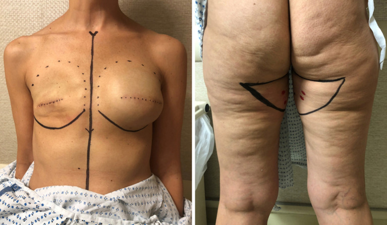
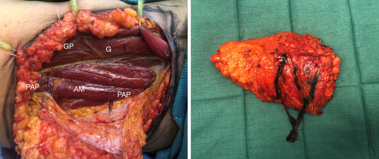
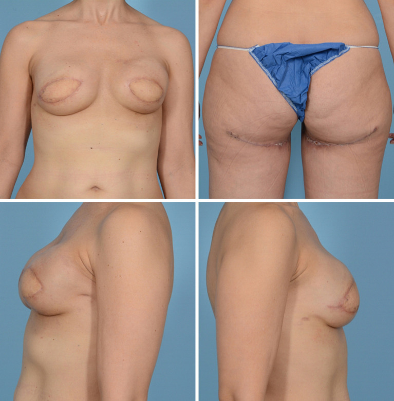
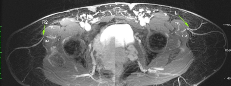
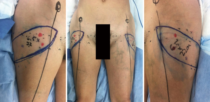
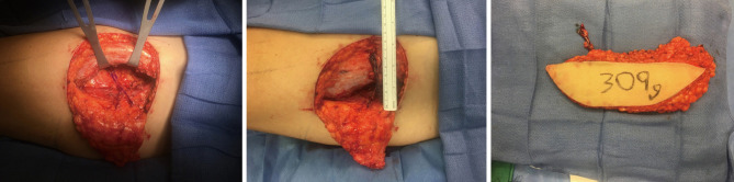
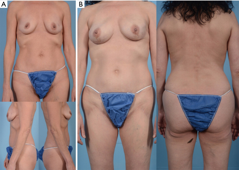
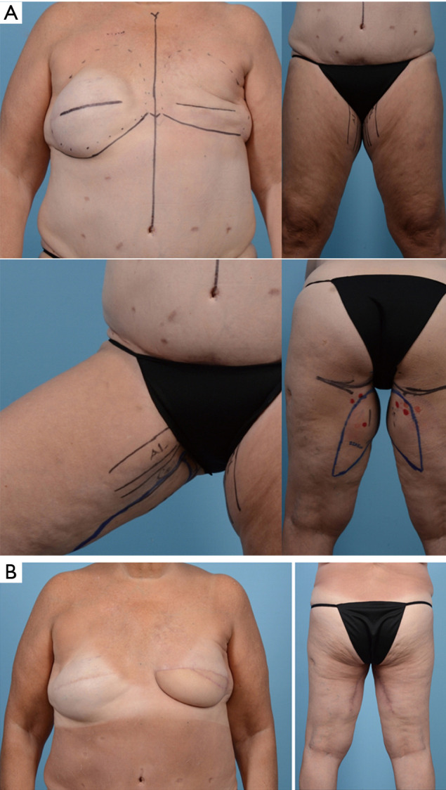
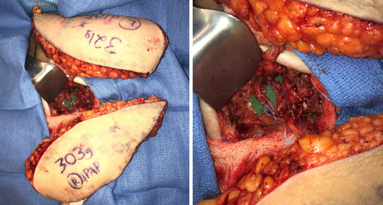
The gold standard for autologous reconstruction in the post-mastectomy patient remains the deep inferior epigastric artery perforator flap, although many women may not be candidates for abdominally based free tissue transfer. In this scenario, there are several other donor site options based from the thigh (transverse and diagonal upper gracilis flaps, profunda artery perforator flap, lateral thigh flap) and trunk (lumbar artery perforator flap, superior and inferior gluteal artery perforator flaps). This study will review the history, relevant anatomy, surgical technique and outcomes for alternative flaps in autologous reconstruction. Additionally, preoperative imaging (CTA, MRA) and novel applications (stacking flaps, neurotization) in breast reconstructive microsurgery will be discussed.

Keywords: Microsurgery; autologous tissue reconstruction; breast reconstruction.


Conflict of interest statement

Conflicts of Interest: All authors have completed the ICMJE uniform disclosure form (available at http://dx.doi.org/10.21037/gs.2020.03.16). The authors have no conflicts of interest to declare.

Figures

Figure 1
Figure 1
MRA showing profunda artery perforator (red arrow). Reference points are the midline, gluteal crease and posterior border of the gracilis muscle. Imaging assists in not only finding the location of the perforator of choice, but also the course it takes to the source vessel. MRA, magnetic resonance angiography.
Figure 2
Figure 2
MRA showing volumetric analysis of left PAP flap (447g for a 6 cm × 22 cm flap). MRA, magnetic resonance angiography.
Figure 3
Figure 3
Preoperative markings for DUG flap. Note the reference points of the adductor longus (AL) and gracilis (G) muscles. DUG, diagonal upper gracilis.
Figure 4
Figure 4
Preoperative (A) and 1 year postoperative (B) photos of DUG flaps for bilateral breast reconstruction.
Figure 5
Figure 5
Preoperative photographs of bilateral PAP flaps for failed right breast tissue expander secondary to infection. The gluteal crease is marked and perforators identified based on preoperative imaging (Figure 1). PAP, profunda artery perforator.
Figure 6
Figure 6
Intraoperative flap dissection of PAP flap. Note the relationship of the PAP to the G, its pedicle and the AM and the flap harvested weighed 430 g as previously predicted on imaging (Figure 2). PAP, profunda artery perforator; GP, pedicle; AM, adductor magnus; G, gracilis muscle.
Figure 7
Figure 7
Postoperative photographs 6 weeks from bilateral PAP flaps. PAP, profunda artery perforator.
Figure 8
Figure 8
Lateral thigh perforator coursing in the posterior septum between the TFL and GM muscles. TFL, tensor fascia latae; GM, gluteus medius.
Figure 9
Figure 9
Lateral thigh perforator flap markings. The pubic symphysis, ASIS, lateral border of the patella and the greater trochanter are identified. A line is drawn that connects the ASIS to the lateral border of the patella, representing the anterior border of the TFL muscle. A second line is drawn laterally from the pubic symphysis intersecting the anterior TFL border. Perforators are identified with Doppler ultrasound in the inferolateral quadrant. Flap width is determined by a pinch test. TFL, tensor fascia latae; ASIS, anterior superior iliac spine.
Figure 10
Figure 10
Intraoperative dissection of LTP flap with division of the pedicle prior to making posterior incision. The flap maintains perfusion through posterior gluteal perforators. LTP, lateral thigh perforator.
Figure 11
Figure 11
Preoperative (A) and 10-month postoperative (B) photos of bilateral LTP flap reconstruction, without revisions. Note the masculinization of the thighs. LTP, lateral thigh perforator.
Figure 12
Figure 12
Preoperative (A) and post-operative (B) photographs of a patient with failure of left breast reconstruction presenting for stacked diagonal PAP flap reconstruction. PAP, profunda artery perforator.
Figure 13
Figure 13
Intraoperative photograph of stacked DPAP flaps with retrograde and anterograde anastomoses. DPAP, diagonal profunda artery perforator.

Similar articles

Cited by

References

    1. Albornoz CR, Bach PB, Mehrara BJ, et al. A paradigm shift in U.S. breast reconstruction: increasing implant rates. Plast Reconstr Surg 2013;131:15-23. 10.1097/PRS.0b013e3182729cde - DOI - PubMed
    1. Avraham T, Clavin N, Mehrara BJ. Microsurgical breast reconstruction. Cancer J 2008;14:241-7 10.1097/PPO.0b013e31817fb7e3 - DOI - PubMed
    1. Yueh JH, Slavin SA, Adesiyun T, et al. Patient satisfaction in postmastectomy breast reconstruction: a comparative evaluation of DIEP, TRAM, latissimus flap, and implant techniques. Plast Reconstr Surg 2010;125:1585-95. 10.1097/PRS.0b013e3181cb6351 - DOI - PubMed
    1. Hartrampf CR, Soneflan M, Black PW. Breast reconstruction with a transverse abdominal island flap. Plast Reconstr Surg 1982;69:216-25. 10.1097/00006534-198202000-00006 - DOI - PubMed
    1. Grotting JC, Urist MM, Maddox WA, et al. Conventional TRAM flap versus free microsurgical TRAM flap for immediate breast reconstruction. Plast Reconstr Surg 1989;83:828-41. 10.1097/00006534-198905000-00009 - DOI - PubMed

LinkOut - more resources