. 2021 Feb 9:10:584024.
doi: 10.3389/fonc.2020.584024. eCollection 2020.

The Cancer/Testis Antigen Gene VCX2 Is Rarely Expressed in Malignancies but Can Be Epigenetically Activated Using DNA Methyltransferase and Histone Deacetylase Inhibitors

Affiliations

The Cancer/Testis Antigen Gene VCX2 Is Rarely Expressed in Malignancies but Can Be Epigenetically Activated Using DNA Methyltransferase and Histone Deacetylase Inhibitors

Mie K Jakobsen et al. Front Oncol. .

Abstract

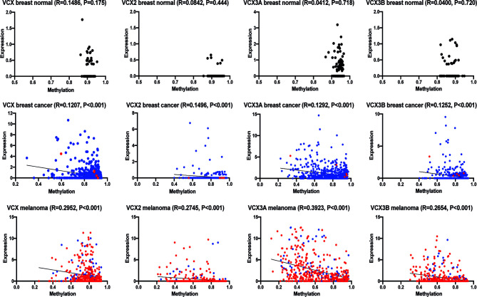
Identification of novel tumor-specific targets is important for the future development of immunotherapeutic strategies using genetically engineered T cells or vaccines. In this study, we characterized the expression of VCX2, a member of the VCX/Y cancer/testis antigen family, in a large panel of normal tissues and tumors from multiple cancer types using immunohistochemical staining and RNA expression data. In normal tissues, VCX2 was detected in the germ cells of the testis at all stages of maturation but not in any somatic tissues. Among malignancies, VCX2 was only found in tumors of a small subset of melanoma patients and thus rarely expressed compared to other cancer/testis antigens such as GAGE and MAGE-A. The expression of VCX2 correlated with that of other VCX/Y genes. Importantly, we found that expression of VCX2 was inversely correlated with promoter methylation and could be activated by treatment with a DNA methyltransferase inhibitor in multiple breast cancer and melanoma cell lines and a breast cancer patient-derived xenograft. The effect could be further potentiated by combining the DNA methyltransferase inhibitor with a histone deacetylase inhibitor. Our results show that the expression of VCX2 can be epigenetically induced in cancer cells and therefore could be an attractive target for immunotherapy of cancer.

Keywords: DNA methyl transferase (DNMT) inhibition; Histone deacetylase inhibitors; Immunotherapy; VCX2; cancer/testis (CT) antigen.


Conflict of interest statement

The authors declare that the research was conducted in the absence of any commercial or financial relationships that could be construed as a potential conflict of interest.

Figures

Figure 1
Figure 1
VCX2 protein expression is limited to testis among healthy adult tissues. (A) The specificity of a VCX2 specific antibody used for immunohistochemical analysis toward VCX2 was confirmed by western blotting using lysates from HEK293T cells with ectopic expression of VCX2 and VCX. The antibody only recognized VCX2 and not VCX or other endogenously expressed proteins. (B) VCX2 protein expression was investigated in a panel of 23 different normal adult tissues. Expression was only observed in testis among these tissues, where nuclear staining was seen in germ cells at all stages of spermatogenesis. Only a subset of tissues is shown. Magnification: × 40.
Figure 2
Figure 2
VCX2 expression in cancer and subcellular localization. (A) VCX2 protein expression was observed in 4/19 melanoma cell lines (FM45, FM79, A375, and Sk-Mel-37b) and 1/11 breast cancer cell lines (T-47-D) (see Supplementary Table 1 for a complete list). (B) The antibody was confirmed to recognize a protein similar in size to VCX2 in melanoma cell lysate from cells positive in immunohistochemical staining (FM45) and did not react with a lysate from cells negative in immunohistochemical staining (FM3). (C) Among 261 cancer specimens derived from 19 different histological origins, VCX2 protein expression was only observed in melanoma, where expression was seen in 4/31 cancer specimens (see Table 1 for a complete list). Only a subset of cancer cell lines and cancer specimens are shown. (D) Investigation of VCX2 subcellular localization in A375 cells, using immunocytochemistry, showed the presence of the protein only in the nucleus, with a localization pattern similar to chromatin. Magnification: × 40 (A, C) × 60 (D).
Figure 3
Figure 3
VCX is expressed in a subset of breast cancer and melanoma tumors and correlates with the expression of other VCX family members. (A) RNA expression of VCX family members in normal breast and breast cancer tissues. Data were generated using RNA sequencing data from the TCGA repository. Results are presented as relative expression levels (normalized number of reads). (B) Expression of VCX family members and GAGE, MAGE-A1, and CTAG1B (NY-ESO-1) CT antigens primary and metastatic melanoma tumors. Data were generated using RNA sequencing data from the TCGA repository. Results are presented as relative expression levels (normalized number of reads). (C) Spearman correlation between expression of VCX2 and other members of the VCX family in primary (blue) and metastatic (red) melanoma tumors. VCX significantly correlated with all other VCX genes. The p-values were calculated using the (unpaired) Mann-Whitney U test.
Figure 4
Figure 4
Correlation of VCX gene promotor methylation levels and mRNA expression. Spearman correlation between average promotor methylation levels, shown as ß-values and mRNA expression of all VCX family genes in healthy and cancerous breast tissue and melanoma. A significant correlation was observed for all VCX family genes in both breast cancer and melanoma, but not in healthy breast tissue. Blue indicates primary tumor specimens and red indicates metastatic tumor specimens. The p-values were calculated using the (unpaired) Mann-Whitney U test.
Figure 5
Figure 5
Upregulation of VCX2 expression by epigenetic treatment in breast cancer and melanoma cells. (A) VCX family gene expression was investigated by RNA sequencing in MDA-MB-231 breast cancer cells after treatment with either vehicle (black bars) or guadecitabine (white bars) for 96 h. All VCX genes were significantly upregulated by treatment with guadecitabine. The experiment was performed with three biological replicates. (B) The effect of combining DNA methyltransferase inhibitors and histone deacetylase inhibitors were investigated using guadecitabine and valproic acid. MDA-MB-231 cells were treated with increasing concentrations of guadecitabine (0.3 to 10 μM) with or without 1 mM valproic acid for 96 h and VCX2 protein expression was subsequently evaluated using immunocytochemical staining. The experiment was performed with two biological replicates. A representative replicate is shown. (C) Quantification of cells from (B). (D) MCF7 breast cancer cells were treated with 0.5 mM of guadecitabine with or without 1 mM valproic acid (VPA) for 96 h and VCX2 protein expression was subsequently evaluated using immunocytochemical staining. The experiment was performed with two biological replicates. A representative replicate is shown. Approximate frequency of positive cells is shown in bottom left corner. (E) PDX tumors from a metastatic TNBC patient were established in NOG mice and when tumors reached a size of 2–3 mm, the mice were randomized into treatment groups based on tumor size. The mice were treated with either 24.4 mg/kg guadecitabine or vehicle every fifth day for a total of four times and VCX2 expression was subsequently evaluated by immunohistochemistry. The experiment was performed with three biological replicates. A representative replicate is shown. Approximate frequency of positive cells is shown in bottom left corner. Scale bars = 50 µM. (F) A375 and MZ2-MEL melanoma cells were treated with 0.5 mM of guadecitabine (gua) with or without 1 mM valproic acid (VPA) for 96 h and VCX2 protein expression was subsequently evaluated using immunocytochemical staining. Representative pictures are shown. Approximate frequency of positive cells is shown in bottom left corner. Scale bars = 50 µM.
Figure 6
Figure 6
VCX2 expression in breast cancer cells is associated with loss of promoter methylation. The level of VCX2 promoter methylation in peripheral blood mononuclear cells (PBMCs) and breast cancer cells lines was investigated using bisulfite conversion followed by PCR melting point analysis. (A) The methylation level in PBMCs was similar to that of in vitro methylated DNA (IVM) and distinct from that of unmethylated DNA prepared by whole genome amplification (WGA). (B) The analysis was performed on cells with (T-47-D) or without (MDA-MB-231) endogenous expression of VCX2 and on cells with guadecitabine/valproic acid-induced expression of VCX2 [MDA-MB-231 + guadecitabine (Gua)/valproic acid (VPA)], which demonstrated inverse correlation between VCX2 promoter methylation and gene expression. Two different promoter regions were analyzed with highly similar results (only data for region 2 are shown). The experiment was performed with two biological replicates.
Figure 7
Figure 7
Proposed model for targeting VCX2 in tumors. VCX2 is rarely expressed in tumors, but can be epigenetically activated by treatment with DNA methyltransferase (DNMT) and histone deacetylase (HDAC) inhibitors. Enhanced production of VCX2 will make tumor cells susceptible to anti-VCX2 T-cell responses or to T-cell therapy with T cells genetically modified to express VCX2-specific T-cell receptors.

Similar articles

Cited by

References

    1. Gjerstorff MF, Andersen MH, Ditzel HJ. Oncogenic cancer/testis antigens: prime candidates for immunotherapy. Oncotarget (2015) 6(18):15772–87. 10.18632/oncotarget.4694 - DOI - PMC - PubMed
    1. Simpson AJ, Caballero OL, Jungbluth A, Chen YT, Old LJ. Cancer/testis antigens, gametogenesis and cancer. Nat Rev Cancer (2005) 5(8):615–25. 10.1038/nrc1669 - DOI - PubMed
    1. Scanlan MJ, Gure AO, Jungbluth AA, Ol LJ, Chen YT. Cancer/testis antigens: an expanding family of targets for cancer immunotherapy. Immunol Rev (2002) 188:22–32. 10.1034/j.1600-065X.2002.18803.x - DOI - PubMed
    1. Greve KB, Pohl M, Olsen KE, Nielsen O, Ditzel HJ, Gjerstorff MF, et al. SSX2-4 expression in early-stage non-small cell lung cancer. Tissue Antigens (2014) 83(5):344–9. 10.1111/tan.12340 - DOI - PubMed
    1. Oshita C, Takikawa M, Kume A, Miyata H, Ashizawa T, Iizuka A, et al. Dendritic cell-based vaccination in metastatic melanoma patients: phase II clinical trial. Oncol Rep (2012) 28(4):1131–8. 10.3892/or.2012.1956 - DOI - PMC - PubMed