Review
. 2021 Feb 9:8:634849.
doi: 10.3389/fcell.2020.634849. eCollection 2020.

Involvement of the Actin Machinery in Programmed Cell Death

Affiliations
Review

Involvement of the Actin Machinery in Programmed Cell Death

Weida Ren et al. Front Cell Dev Biol. .

Abstract

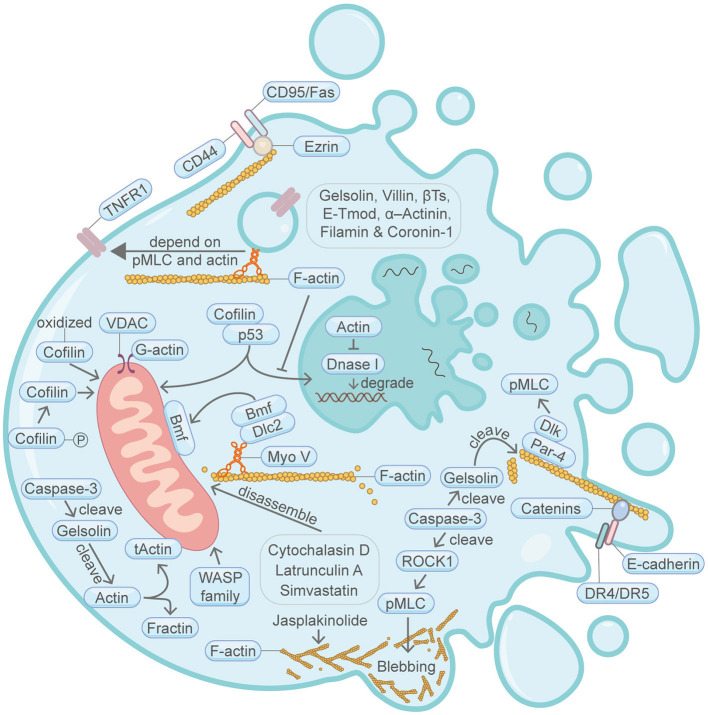
Programmed cell death (PCD) depicts a genetically encoded and an orderly mode of cellular mortality. When triggered by internal or external stimuli, cells initiate PCDs through evolutionary conserved regulatory mechanisms. Actin, as a multifunctional cytoskeleton protein that forms microfilament, its integrity and dynamics are essential for a variety of cellular processes (e.g., morphogenesis, membrane blebbing and intracellular transport). Decades of work have broadened our knowledge about different types of PCDs and their distinguished signaling pathways. However, an ever-increasing pool of evidences indicate that the delicate relationship between PCDs and the actin cytoskeleton is beginning to be elucidated. The purpose of this article is to review the current understanding of the relationships between different PCDs and the actin machinery (actin, actin-binding proteins and proteins involved in different actin signaling pathways), in the hope that this attempt can shed light on ensuing studies and the development of new therapeutic strategies.

Keywords: actin; actin machinery; actin-binding proteins; actin-modulating proteins; cytoskeleton; programmed cell death.


Conflict of interest statement

The authors declare that the research was conducted in the absence of any commercial or financial relationships that could be construed as a potential conflict of interest.

Figures

Figure 1
Figure 1
Schematic illustrating apoptosis and the actin machinery. Actin-binding proteins, such as Gelsolin, Villin, β-Thymosins, E-tropomodulin, Filamin and Coronin-1, play active roles in apoptosis. The actin cytoskeleton integrity is essential for CD95/Fas-mediated apoptosis. During another TNF-induced apoptosis, plasma membrane translocation of the TNFR1 requires myosin II motor and actin. E-cadherin and catenins engagement can augment apoptosis activation by linking DR4/DR5 to the F-actin cytoskeleton. Bmf translocation from the filamentous actin to the mitochondria is important for apoptosis. Cofilin protein amount and its posttranslational modification status are important for apoptosis. Cytochalasin D, latrunculin A and Simvastatin can induce apoptosis through disrupting the actin cytoskeleton network. Cofilin and actin affects p53-mediated control of apoptosis. Par-4 can recruit Dlk to the filamentous actin, thereby enhancing the phosphorylation of MLC and induction of apoptosis. WASP family protein WAVE1 can regulate apoptosis through affecting mitochondria. Actin is cleaved by caspase during apoptosis, resulting in the production of tActin and Fractin. tActin, rather than Fractin, can specifically induce morphological changes resembling apoptosis. Actin is also involved in the regulation of DNA degradation during apoptosis. Membrane blebbing is supervised by actomyosin contractility, which in turn is regulated by Caspase-3-ROCK1 cleavage-pMLC axis. Please see the main text for more detailed information. Abbreviations: Caspase, cysteine aspartic protease; βTs, β-Thymosins; E-Tmod, E-tropomodulin; TNFR1, TNF receptor-1; VDAC, voltage-dependent anion channel; Bmf, Bcl2-modifying factor; Dlc2, dynein light chain 2; pMLC, phosphorylated myosin light chain; Dlk, DAP like kinase; Par-4, prostate apoptosis response-4; DR4/DR5, death receptor 4/5; ROCK1, Rho-associated coiled-coil kinase; Myo V, myosin V; WASP, Wiskott-Aldrich Syndrome protein; tActin, mitochondria-targeted N-myristoylated 15 kDa fragment of actin; Fractin, N-terminal 32 kDa fragment of actin; Dnase I, deoxyribonuclease I.
Figure 2
Figure 2
Schematic illustrating lysosomal cell death and the actin machinery. Permeabilization of lysosomal membrane and the subsequent release of hydrolytic enzymes including cathepsins are key features of lysosomal cell death. Direct evidence connecting lysosomal cell death and the actin machinery is limited. Nevertheless, certain proteins of the actin machinery, such as actin, myosin and Cofilin, can be degraded or modulated by cathepsins. Lysosome movement also closely correlates with F-actin and myosin. GA101, a type II CD20-targeted monoclonal antibody, can induce lysosomal cell death. This GA101-induced cell death can be abrogated by inhibitors of actin polymerization. Please see the main text for more detailed information. Abbreviations: LMP, lysosomal membrane permeabilization; MHC, myosin heavy chain.
Figure 3
Figure 3
Schematic illustrating pyroptosis and the actin machinery. Actin machinery is required for pyroptosis. F-actin forms around phagosomes at the early stage of pyroptosis. Cortical actin networks are disrupted during the progression of pyroptosis. Caspase-11 and RhoA maintain the actin-severing protein Cofilin in the phosphorylated inactive form, which sustains actin polymerization and mediates phagosome-lysosome fusion. Cofilin activity is also regulated by Caspase-1 and Slingshot. NLRP3 inflammasome also represses F-actin remodeling. The ESCRT-III complex repairs damaged plasma membrane. ESCRT-III cooperates with the actin cytoskeleton during other processes such as cytokinesis and wound healing. Please see the main text for more detailed information. Abbreviations: GSDMD, gasdermin D; ESCRT, endosomal sorting complexes required for transport; NLRP3, NLR family pyrin domain containing 3; RhoA, Ras homolog family member A; Caspase, cysteine–aspartate protease.
Figure 4
Figure 4
Schematic illustrating NETosis and the actin machinery. NETosis is exclusively found in neutrophils. Cortical F-actin disassembly is a prerequisite for NET release and occurs at the early stage of NETosis. Neutrophil elastase can bind and degrade F-actin network. Both F-actin stabilization by jasplakinolide and F-actin depolymerization by cytochalasin D lead to the attenuation of NET release. Execution of PKCα function in the nucleus is important for nuclear envelope rupture and NET formation. PKCα nuclear translocation requires intact actin cytoskeleton in NIH 3T3 fibroblasts. Please see the main text for more detailed information. Abbreviations: NET, neutrophil extracellular traps; PKCα, protein kinase C alpha.
Figure 5
Figure 5
Schematic illustrating necroptosis and the actin machinery. MLKL is the terminal protein in necroptosis. During MLKL translocation to the plasma membrane, it co-traffics with tight junction proteins through Golgi-microtubule-actin-dependent mechanisms. The ESCRT-III complex, which coordinates with the actin cytoskeleton in other contexts, repairs damaged plasma membrane. Please see the main text for more detailed information. Abbreviations: MLKL, mixed lineage kinase domain-like pseudokinase; ESCRT, endosomal sorting complexes required for transport.
Figure 6
Figure 6
Schematic illustrating entosis and the actin machinery. Actin machinery is essential for entosis. In the loser cell, signaling pathway proteins including Cdc42, RhoA, ROCK I/II, Rac1, mDia1, AMPK, PCDH7, PP1α, MRTF, SRF, and Ezrin converge on the actomyosin network to regulate entosis. In the winner cell, PCDH7 and PP1α also exist. Three actin-correlated structures, the contractile actomyosin, the adherens junction and the mechanical ring, are sandwiched between the winner cell and the loser cell. Please see the main text for more detailed information. Abbreviations: Cdc42, cell division control protein 42; RhoA, Ras homolog family member A; ROCK, Rho-associated coiled-coil kinase; Rac1, Ras-related C3 botulinum toxin substrate 1; pMLC2, phosphorylated myosin light chain 2; AMPK, AMP-activated protein kinase; mDia1, diaphanous-related formin 1; PCDH7, protocadherin-7; PP1α, protein phosphatase 1α; MRTF, myocardin-related transcription factor; SRF, serum response factor; CA, the contractile actomyosin; MR, the mechanical ring; AJ, the adherens junction.
Figure 7
Figure 7
Schematic illustrating parthanatos and the actin machinery. It is unclear whether the actin machinery is directly involved in parthanatos. MIF and PARP family proteins are found to affect the actin cytoskeleton in other contexts. Please see the main text for more detailed information. Abbreviations: PAR, poly(ADP-ribose); PARP-1, poly(ADP-ribose) polymerase 1; MIF, migration inhibitory factor; AIF, apoptosis-inducing factor; NAD+, nicotinamide adenine dinucleotide; PARG, poly(ADP-ribose) glycohydrolase.
Figure 8
Figure 8
Schematic illustrating ferroptosis and the actin machinery. PKC-HSPB1 attenuates ferroptosis. HSPB1 regulates F-actin cytoskeleton and TFR1 in other contexts. Cytochalasin D, which depolymerize F-actin, promotes ferroptosis. Suppression of WAVE2, which regulates the branched F-actin network assembly in other studies, favors ferroptosis. Nrf2, p53, and VDAC may be modulated by actin. The ESCRT-III complex, which coordinates with the actin cytoskeleton in other contexts, repairs damaged plasma membrane. Please see the main text for more detailed information. Abbreviations: HSPB1, heat shock protein beta-1; PKC, protein kinase C; WAVE2, Wiskott-Aldrich Syndrome protein family member 2; Keap1, Kelch-like ECH-associated protein 1; VDAC, voltage-dependent anion channel; ESCRT, endosomal sorting complexes required for transport.
Figure 9
Figure 9
Schematic illustrating autosis and the actin machinery. It is unclear whether the actin machinery is directly involved in autosis. In other contexts, Na+,K+-ATPase is found to interact with actin, Cofilin and multiple myosin motors. Na+,K+-ATPase may also be affected by the actin cytoskeleton through α-adducin. Please see the main text for more detailed information. Symbols: K+, potassium ion; Na+, sodium ion.
Figure 10
Figure 10
Schematic illustrating oxeiptosis and the actin machinery. It is unclear whether the actin machinery is directly involved in oxeiptosis. Keap1, an essential protein in oxeiptosis, highly correlates with the actin machinery in other contexts. Please see the main text for more detailed information. Abbreviations: Keap1, Kelch-like ECH-associated protein 1; PGAM5, PGAM family member 5; AIFM1, apoptosis-inducing factor mitochondria associated 1.

Similar articles

Cited by

References

    1. Abu Khweek A., Amer A. O. (2020). Pyroptotic and non-pyroptotic effector functions of caspase-11. Immunol. Rev. 297, 39–52. 10.1111/imr.12910 - DOI - PMC - PubMed
    1. Aida N., Ushikubo T., Kobayashi F., Sako R., Suehara M., Furusawa M., et al. . (2016). Actin stabilization induces apoptosis in cultured porcine epithelial cell rests of Malassez. Int. Endod. J. 49, 663–669. 10.1111/iej.12494 - DOI - PubMed
    1. Akhter A., Caution K., Abu Khweek A., Tazi M., Abdulrahman B. A., Abdelaziz D. H., et al. . (2012). Caspase-11 promotes the fusion of phagosomes harboring pathogenic bacteria with lysosomes by modulating actin polymerization. Immunity 37, 35–47. 10.1016/j.immuni.2012.05.001 - DOI - PMC - PubMed
    1. Albrengues J., Wysocki R. W., Maiorino L., Egeblad M. (2017). Re-cyclin' cell-cycle components to make NETs. Dev. Cell 43, 379–380. 10.1016/j.devcel.2017.11.002 - DOI - PubMed
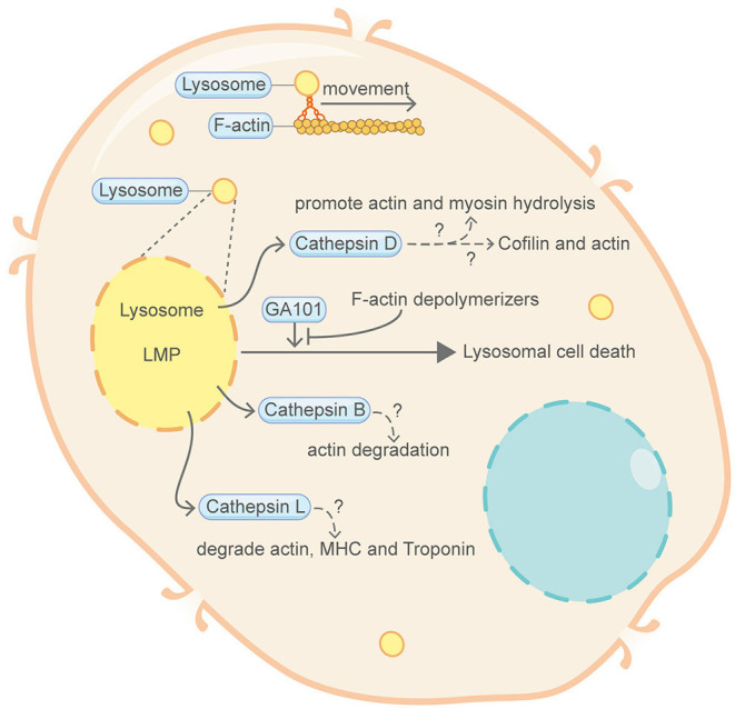
    1. Alduaij W., Ivanov A., Honeychurch J., Cheadle E. J., Potluri S., Lim S. H., et al. . (2011). Novel type II anti-CD20 monoclonal antibody (GA101) evokes homotypic adhesion and actin-dependent, lysosome-mediated cell death in B-cell malignancies. Blood 117, 4519–4529. 10.1182/blood-2010-07-296913 - DOI - PMC - PubMed

LinkOut - more resources