Acyl Transfer Catalytic Activity in De Novo Designed Protein with N-Terminus of α-Helix As Oxyanion-Binding Site
- PMID: 33635059
- PMCID: PMC8012002
- DOI: 10.1021/jacs.0c10053
Acyl Transfer Catalytic Activity in De Novo Designed Protein with N-Terminus of α-Helix As Oxyanion-Binding Site
Abstract
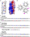
The design of catalytic proteins with functional sites capable of specific chemistry is gaining momentum and a number of artificial enzymes have recently been reported, including hydrolases, oxidoreductases, retro-aldolases, and others. Our goal is to develop a peptide ligase for robust catalysis of amide bond formation that possesses no stringent restrictions to the amino acid composition at the ligation junction. We report here the successful completion of the first step in this long-term project by building a completely de novo protein with predefined acyl transfer catalytic activity. We applied a minimalist approach to rationally design an oxyanion hole within a small cavity that contains an adjacent thiol nucleophile. The N-terminus of the α-helix with unpaired hydrogen-bond donors was exploited as a structural motif to stabilize negatively charged tetrahedral intermediates in nucleophilic addition-elimination reactions at the acyl group. Cysteine acting as a principal catalytic residue was introduced at the second residue position of the α-helix N-terminus in a designed three-α-helix protein based on structural informatics prediction. We showed that this minimal set of functional elements is sufficient for the emergence of catalytic activity in a de novo protein. Using peptide-αthioesters as acyl-donors, we demonstrated their catalyzed amidation concomitant with hydrolysis and proved that the environment at the catalytic site critically influences the reaction outcome. These results represent a promising starting point for the development of efficient catalysts for protein labeling, conjugation, and peptide ligation.
Figures


Similar articles
-
Systemic pharmacological treatments for chronic plaque psoriasis: a network meta-analysis.Cochrane Database Syst Rev. 2021 Apr 19;4(4):CD011535. doi: 10.1002/14651858.CD011535.pub4. Cochrane Database Syst Rev. 2021. Update in: Cochrane Database Syst Rev. 2022 May 23;5:CD011535. doi: 10.1002/14651858.CD011535.pub5. PMID: 33871055 Free PMC article. Updated.
-
Systemic pharmacological treatments for chronic plaque psoriasis: a network meta-analysis.Cochrane Database Syst Rev. 2017 Dec 22;12(12):CD011535. doi: 10.1002/14651858.CD011535.pub2. Cochrane Database Syst Rev. 2017. Update in: Cochrane Database Syst Rev. 2020 Jan 9;1:CD011535. doi: 10.1002/14651858.CD011535.pub3. PMID: 29271481 Free PMC article. Updated.
-
Signs and symptoms to determine if a patient presenting in primary care or hospital outpatient settings has COVID-19.Cochrane Database Syst Rev. 2022 May 20;5(5):CD013665. doi: 10.1002/14651858.CD013665.pub3. Cochrane Database Syst Rev. 2022. PMID: 35593186 Free PMC article.
-
The Black Book of Psychotropic Dosing and Monitoring.Psychopharmacol Bull. 2024 Jul 8;54(3):8-59. Psychopharmacol Bull. 2024. PMID: 38993656 Free PMC article. Review.
-
Comparison of Two Modern Survival Prediction Tools, SORG-MLA and METSSS, in Patients With Symptomatic Long-bone Metastases Who Underwent Local Treatment With Surgery Followed by Radiotherapy and With Radiotherapy Alone.Clin Orthop Relat Res. 2024 Dec 1;482(12):2193-2208. doi: 10.1097/CORR.0000000000003185. Epub 2024 Jul 23. Clin Orthop Relat Res. 2024. PMID: 39051924
Cited by
-
From peptides to proteins: coiled-coil tetramers to single-chain 4-helix bundles.Chem Sci. 2022 Sep 20;13(38):11330-11340. doi: 10.1039/d2sc04479j. eCollection 2022 Oct 5. Chem Sci. 2022. PMID: 36320580 Free PMC article.
-
Sparks of function by de novo protein design.Nat Biotechnol. 2024 Feb;42(2):203-215. doi: 10.1038/s41587-024-02133-2. Epub 2024 Feb 15. Nat Biotechnol. 2024. PMID: 38361073 Free PMC article. Review.
-
Site-selective peptide bond hydrolysis and ligation in water by short peptide-based assemblies.Proc Natl Acad Sci U S A. 2024 Jul 30;121(31):e2321396121. doi: 10.1073/pnas.2321396121. Epub 2024 Jul 23. Proc Natl Acad Sci U S A. 2024. PMID: 39042686 Free PMC article.
-
Biocatalysts Based on Peptide and Peptide Conjugate Nanostructures.Biomacromolecules. 2021 May 10;22(5):1835-1855. doi: 10.1021/acs.biomac.1c00240. Epub 2021 Apr 12. Biomacromolecules. 2021. PMID: 33843196 Free PMC article. Review.
-
Design and Engineering of Miniproteins.ACS Bio Med Chem Au. 2022 Apr 28;2(4):316-327. doi: 10.1021/acsbiomedchemau.2c00008. eCollection 2022 Aug 17. ACS Bio Med Chem Au. 2022. PMID: 37102166 Free PMC article. Review.
References
-
- Zanghellini A De Novo Computational Enzyme Design. Curr. Opin. Biotechnol 2014, 29, 132–138. - PubMed
-
- Kries H; Blomberg R; Hilvert D De Novo Enzymes by Computational Design. Curr. Opin. Chem. Biol 2013, 17, 221–228. - PubMed
-
- Hilvert D Design of Protein Catalysts. Annu. Rev. Biochem 2013, 82, 447–470. - PubMed
-
- Röthlisberger D; Khersonsky O; Wollacott AM; Jiang L; DeChancie J; Betker J; Gallaher JL; Althoff EA; Zanghellini A; Dym O; Albeck S; Houk KN; Tawfik DS; Baker D Kemp Elimination Catalysts by Computational Enzyme Design. Nature 2008, 453, 190–195. - PubMed
Publication types
MeSH terms
Substances
Grants and funding
LinkOut - more resources
Full Text Sources
Other Literature Sources
Research Materials

