What Is the Comparative Ability of 18F-FDG PET/CT, 99mTc-MDP Skeletal Scintigraphy, and Whole-body MRI as a Staging Investigation to Detect Skeletal Metastases in Patients with Osteosarcoma and Ewing Sarcoma?
- PMID: 33635285
- PMCID: PMC8277296
- DOI: 10.1097/CORR.0000000000001681
What Is the Comparative Ability of 18F-FDG PET/CT, 99mTc-MDP Skeletal Scintigraphy, and Whole-body MRI as a Staging Investigation to Detect Skeletal Metastases in Patients with Osteosarcoma and Ewing Sarcoma?
Abstract
Background: Skeletal metastases of bone sarcomas are indicators of poor prognosis. Various imaging modalities are available for their identification, which include bone scan, positron emission tomography/CT scan, MRI, and bone marrow aspiration/biopsy. However, there is considerable ambiguity regarding the best imaging modality to detect skeletal metastases. To date, we are not sure which of these investigations is best for screening of skeletal metastasis.
Question/purpose: Which staging investigation-18F-fluorodeoxyglucose positron emission tomography/CT (18F-FDG PET/CT), whole-body MRI, or 99mTc-MDP skeletal scintigraphy-is best in terms of sensitivity, specificity, positive predictive value (PPV), and negative predictive value (NPV) in detecting skeletal metastases in patients with osteosarcoma and those with Ewing sarcoma?
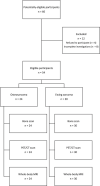
Methods: A prospective diagnostic study was performed among 54 of a total 66 consecutive osteosarcoma and Ewing sarcoma patients who presented between March 2018 and June 2019. The institutional review board approved the use of all three imaging modalities on each patient recruited for the study. Informed consent was obtained after thoroughly explaining the study to the patient or the patient's parent/guardian. The patients were aged between 4 and 37 years, and their diagnoses were proven by histopathology. All patients underwent 99mTc-MDP skeletal scintigraphy, 18F-FDG PET/CT, and whole-body MRI for the initial staging of skeletal metastases. The number and location of bone and bone marrow lesions diagnosed with each imaging modality were determined and compared with each other. Multidisciplinary team meetings were held to reach a consensus about the total number of metastases present in each patient, and this was considered the gold standard. The sensitivity, specificity, PPV, and NPV of each imaging modality, along with their 95% confidence intervals, were generated by the software Stata SE v 15.1. Six of 24 patients in the osteosarcoma group had skeletal metastases, as did 8 of 30 patients in the Ewing sarcoma group. The median (range) follow-up for the study was 17 months (12 to 27 months). Although seven patients died before completing the minimum follow-up, no patients who survived were lost to follow-up.
Results: With the number of patients available, we found no differences in terms of sensitivity, specificity, PPV, and NPV among the three staging investigations in patients with osteosarcoma and in patients with Ewing sarcoma. Sensitivities to detect bone metastases for 18F-FDG PET/CT, whole-body MRI, and 99mTc-MDP skeletal scintigraphy were 100% (6 of 6 [95% CI 54% to 100%]), 83% (5 of 6 [95% CI 36% to 100%]), and 67% (4 of 6 [95% CI 22% to 96%]) and specificities were 100% (18 of 18 [95% CI 82% to 100%]), 94% (17 of 18 [95% CI 73% to 100%]), and 78% (14 of 18 [95% CI 52% to 94%]), respectively, in patients with osteosarcoma. In patients with Ewing sarcoma, sensitivities to detect bone metastases for 18F-FDG PET/CT, whole-body MRI, and 99mTc-MDP skeletal scintigraphy were 88% (7 of 8 [95% CI 47% to 100%]), 88% (7 of 8 [95% CI 47% to 100%]), and 50% (4 of 8 [95% CI 16% to 84%]) and specificities were 100% (22 of 22 [95% CI 85% to 100%]), 95% (21 of 22 [95% CI 77% to 100%]), and 95% (21 of 22 [95% CI 77% to 100%]), respectively. Further, the PPVs for detecting bone metastases for 18F-FDG PET/CT, whole-body MRI, and 99mTc-MDP skeletal scintigraphy were 100% (6 of 6 [95% CI 54% to 100%]), 83% (5 of 6 [95% CI 36% to 100%]), and 50% (4 of 8 [95% CI 16% to 84%]) and the NPVs were 100% (18 of 18 [95% CI 82% to 100%]), 94% (17 of 18 [95% CI 73% to 100%]), and 88% (14 of 16 [95% CI 62% to 98%]), respectively, in patients with osteosarcoma. Similarly, the PPVs for detecting bone metastases for 18F-FDG PET/CT, whole-body MRI, and 99mTc-MDP skeletal scintigraphy were 100% (7 of 7 [95% CI 59% to 100%]), 88% (7 of 8 [95% CI 50% to 98%]), and 80% (4 of 5 [95% CI 28% to 100%]), and the NPVs were 96% (22 of 23 [95% CI 78% to 100%]), 95% (21 of 22 [95% CI 77% to 99%]), and 84% (21 of 25 [95% CI 64% to 96%]), respectively, in patients with Ewing sarcoma. The confidence intervals around these values overlapped with each other, thus indicating no difference between them.
Conclusion: Based on these results, we could not demonstrate a difference in the sensitivity, specificity, PPV, and NPV between 18F-FDG PET/CT, whole-body MRI, and 99mTc-MDP skeletal scintigraphy for detecting skeletal metastases in patients with osteosarcoma and Ewing sarcoma. For proper prognostication, a thorough metastatic workup is essential, which should include a highly sensitive investigation tool to detect skeletal metastases. However, our study findings suggest that there is no difference between these three imaging tools. Since this is a small group of patients in whom it is difficult to make broad recommendations, these findings may be confirmed by larger studies in the future.
Level of evidence: Level II, diagnostic study.
Copyright © 2021 by the Association of Bone and Joint Surgeons.
Conflict of interest statement
All ICMJE Conflict of Interest Forms for authors and Clinical Orthopaedics and Related Research® editors and board members are on file with the publication and can be viewed on request. Each author certifies that neither he, nor any member of his immediate family, has funding or commercial associations (consultancies, stock ownership, equity interest, patent/licensing arrangements, etc.) that might pose a conflict of interest in connection with the submitted article.
Figures


Similar articles
-
18F-FDG PET-CT versus MRI for detection of skeletal metastasis in Ewing sarcoma.Skeletal Radiol. 2019 Nov;48(11):1735-1746. doi: 10.1007/s00256-019-03192-2. Epub 2019 Apr 23. Skeletal Radiol. 2019. PMID: 31016339 Free PMC article.
-
Diagnostic performance of 18F-FDG PET/CT and whole-body diffusion-weighted imaging with background body suppression (DWIBS) in detection of lymph node and bone metastases from pediatric neuroblastoma.Ann Nucl Med. 2018 Jun;32(5):348-362. doi: 10.1007/s12149-018-1254-z. Epub 2018 Apr 17. Ann Nucl Med. 2018. PMID: 29667143 Free PMC article.
-
Prospective Comparison of 99mTc-MDP Scintigraphy, Combined 18F-NaF and 18F-FDG PET/CT, and Whole-Body MRI in Patients with Breast and Prostate Cancer.J Nucl Med. 2015 Dec;56(12):1862-8. doi: 10.2967/jnumed.115.162610. Epub 2015 Sep 24. J Nucl Med. 2015. PMID: 26405167
-
The Utility of 18FDG PET/CT Versus Bone Scan for Identification of Bone Metastases in a Pediatric Sarcoma Population and a Review of the Literature.J Pediatr Hematol Oncol. 2021 Mar 1;43(2):52-58. doi: 10.1097/MPH.0000000000001917. J Pediatr Hematol Oncol. 2021. PMID: 32815877 Review.
-
18F-FDG PET/CT and whole-body MRI diagnostic performance in M staging for non-small cell lung cancer: a systematic review and meta-analysis.Eur Radiol. 2020 Jul;30(7):3641-3649. doi: 10.1007/s00330-020-06703-1. Epub 2020 Mar 3. Eur Radiol. 2020. PMID: 32125513
Cited by
-
Advances in the Molecular Imaging of Sarcoma: An Emphasis on Metabolic Imaging.Mol Imaging Biol. 2025 Aug 19. doi: 10.1007/s11307-025-02045-w. Online ahead of print. Mol Imaging Biol. 2025. PMID: 40830327 Review.
-
PET-CT in Clinical Adult Oncology-VI. Primary Cutaneous Cancer, Sarcomas and Neuroendocrine Tumors.Cancers (Basel). 2022 Jun 8;14(12):2835. doi: 10.3390/cancers14122835. Cancers (Basel). 2022. PMID: 35740501 Free PMC article. Review.
-
Multimodal Imaging of Osteosarcoma: From First Diagnosis to Radiomics.Cancers (Basel). 2025 Feb 10;17(4):599. doi: 10.3390/cancers17040599. Cancers (Basel). 2025. PMID: 40002194 Free PMC article. Review.
-
Effect of Paclitaxel Combined with Doxorubicin Hydrochloride Liposome Injection in the Treatment of Osteosarcoma and MRI Changes before and after Treatment.Evid Based Complement Alternat Med. 2022 Jul 30;2022:5651793. doi: 10.1155/2022/5651793. eCollection 2022. Evid Based Complement Alternat Med. 2022. Retraction in: Evid Based Complement Alternat Med. 2023 Jul 19;2023:9816837. doi: 10.1155/2023/9816837. PMID: 35942377 Free PMC article. Retracted.
-
Ewing sarcoma of the temporal bone with aneurysmal bone cyst-like changes: A rare case report with an unusual radiological presentation.Neuroradiol J. 2024 Oct;37(5):640-644. doi: 10.1177/19714009231212358. Epub 2023 Nov 3. Neuroradiol J. 2024. PMID: 37923348
References
-
- Anninga JK, Gelderblom H, Fiocco M, et al. Chemotherapeutic adjuvant treatment for osteosarcoma: where do we stand? Eur J Cancer. 2011;47:2431-2445. - PubMed
-
- Connolly LP, Drubach LA, Treves TS. Applications of nuclear medicine in pediatric oncology. Clin Nucl Med. 2002;27:117-125. - PubMed
-
- Daldrup-Link HE, Franzius C, Link TM, et al. Whole-body MR imaging for detection of bone metastases in children and young adults: comparison with skeletal scintigraphy and FDG PET. Am J Roentgenol. 2001;177:229-236. - PubMed
-
- Dean BJ, Whitwell D. Epidemiology of bone and soft-tissue sarcomas. Orthop Trauma. 2009;23:223-230.
-
- Franzius C, Sciuk J, Daldrup-Link HE, Jürgens H, Schober O. FDG-PET for detection of osseous metastases from malignant primary bone tumours: comparison with bone scintigraphy. Eur J Nucl Med. 2000;27:1305-1311. - PubMed
Publication types
MeSH terms
Substances
LinkOut - more resources
Full Text Sources
Other Literature Sources
Medical
Research Materials

