

Descriptive analysis of a comparison between lung ultrasound and chest radiography in patients suspected of COVID-19
- PMID: 33635443
- PMCID: PMC7907795
- DOI: 10.1186/s13089-021-00215-9
Descriptive analysis of a comparison between lung ultrasound and chest radiography in patients suspected of COVID-19
Abstract
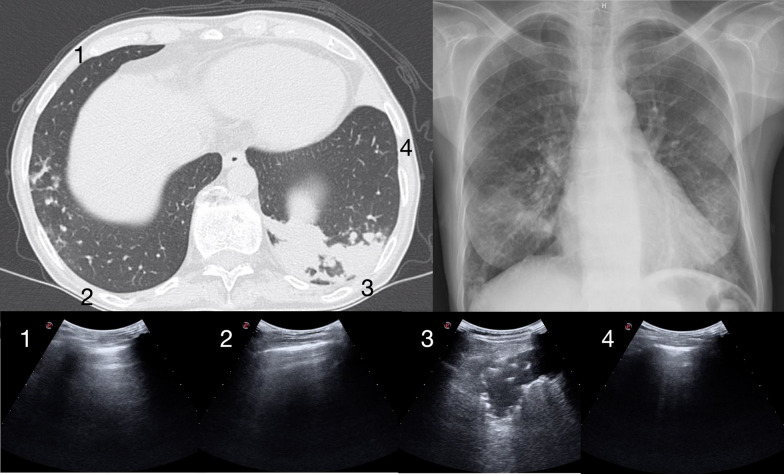
Background: Lung ultrasound (LUS) and chest radiography (CXR) are the most used chest imaging tools in the early diagnosis of COVID-19 associated pneumonia. However, the relationship between LUS and CXR is not clearly defined. The aim of our study was to describe the comparison between LUS interpretation and CXR readings in the first approach to patients suspected of COVID-19.
Methods: In the time of the first COVID-19 pandemic surge, we prospectively evaluated adult patients presenting to an emergency department complaining of symptoms raising suspicion of COVID-19. Patients were studied by LUS and only those performing also CXR were analyzed. All the patients performed viral reverse transcriptase-polymerase chain reaction (RT-PCR). LUS studies were classified in 4 categories of probabilities, based on the presence of typical or alternative signs of COVID-19-associated interstitial pneumonia. Accordingly, the CXR readings were retrospectively adapted by 2 experts in 4 categories following the standard language that describes the computed tomography (CT) findings. Patients were divided in two groups, based on the agreement of the LUS and CXR categories. Results were also compared to RT-PCR and, when available, to CT studies.
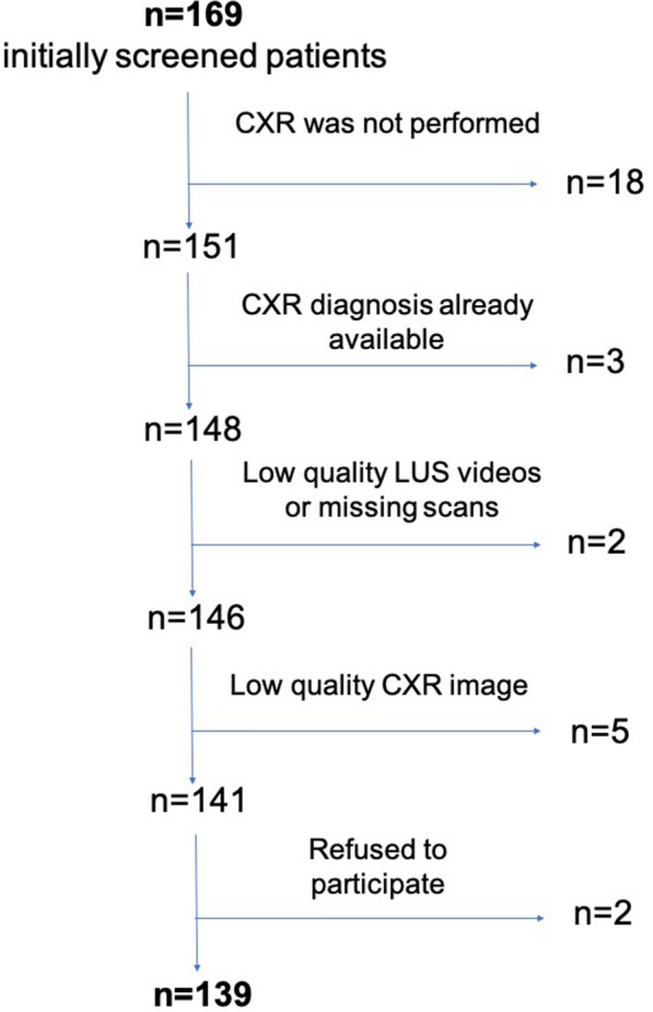
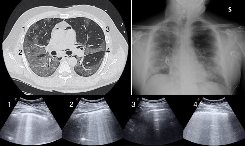
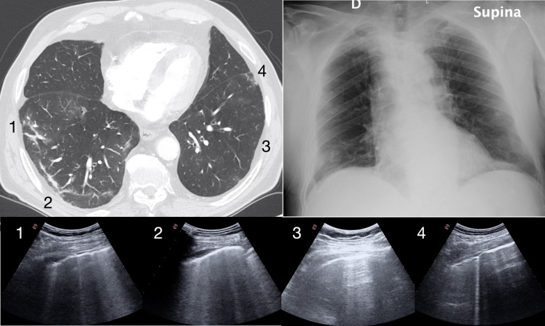
Results: We analyzed 139 cases (55 women, mean age 59.1 ± 15.5 years old). The LUS vs CXR results disagreed in 60 (43.2%) cases. RT-PCR was positive in 88 (63.3%) cases. In 45 cases, a CT scan was also performed and only 4 disagreed with LUS interpretation versus 24 in the comparison between CT and CXR. In 18 cases, LUS detected signs of COVID-19 pneumonia (high and intermediate probabilities) while CXR reading was negative; in 14 of these cases, a CT scan or a RT-PCR-positive result confirmed the LUS interpretation. In 6 cases, LUS detected signs of alternative diagnoses to COVID-19 pneumonia while CXR was negative; in 4 of these cases, CT scan confirmed atypical findings.
Conclusion: Our study demonstrated a strong disagreement between LUS interpretation and CXR reading in the early approach to patients suspected of COVID-19. Comparison with CT studies and RT-PCR results seems to confirm the superiority of LUS over a second retrospective reading of CXR.
Conflict of interest statement
The authors declare that they have no competing interests with the subject of the article.
Figures
Similar articles
-
Lung Ultrasound vs. Chest X-Ray Study for the Radiographic Diagnosis of COVID-19 Pneumonia in a High-Prevalence Population.J Emerg Med. 2021 May;60(5):615-625. doi: 10.1016/j.jemermed.2021.01.041. Epub 2021 Feb 4. J Emerg Med. 2021. PMID: 33722414 Free PMC article.
-
Comparison of Lung Ultrasound versus Chest X-ray for Detection of Pulmonary Infiltrates in COVID-19.Diagnostics (Basel). 2021 Feb 22;11(2):373. doi: 10.3390/diagnostics11020373. Diagnostics (Basel). 2021. PMID: 33671699 Free PMC article.
-
Lung Ultrasonography: A Viable Alternative to Chest Radiography in Children with Suspected Pneumonia?J Pediatr. 2016 Sep;176:93-98.e7. doi: 10.1016/j.jpeds.2016.05.033. Epub 2016 Jun 16. J Pediatr. 2016. PMID: 27318374
-
Thoracic imaging tests for the diagnosis of COVID-19.Cochrane Database Syst Rev. 2020 Sep 30;9:CD013639. doi: 10.1002/14651858.CD013639.pub2. Cochrane Database Syst Rev. 2020. Update in: Cochrane Database Syst Rev. 2020 Nov 26;11:CD013639. doi: 10.1002/14651858.CD013639.pub3. PMID: 32997361 Updated.
-
The utility of chest x-ray and lung ultrasound in the management of infants and children presenting with severe pneumonia in low-and middle-income countries: A pragmatic scoping review.J Glob Health. 2022 Dec 23;12:10013. doi: 10.7189/jogh.12.10013. J Glob Health. 2022. PMID: 36560909 Free PMC article.
Cited by
-
Assessing COVID-19 pneumonia-Clinical extension and risk with point-of-care ultrasound: A multicenter, prospective, observational study.J Am Coll Emerg Physicians Open. 2021 May 1;2(3):e12429. doi: 10.1002/emp2.12429. eCollection 2021 Jun. J Am Coll Emerg Physicians Open. 2021. PMID: 33969350 Free PMC article.
-
Point-of-care ultrasound use in COVID-19: a narrative review.Ann Transl Med. 2024 Feb 1;12(1):13. doi: 10.21037/atm-23-1403. Epub 2023 Jul 13. Ann Transl Med. 2024. PMID: 38304913 Free PMC article. Review.
-
Ten Questions on Using Lung Ultrasonography to Diagnose and Manage Pneumonia in the Hospital-at-Home Model: Part I-Techniques and Patterns.Diagnostics (Basel). 2024 Dec 13;14(24):2799. doi: 10.3390/diagnostics14242799. Diagnostics (Basel). 2024. PMID: 39767160 Free PMC article. Review.
-
Implementing Lung Ultrasound in the Outpatient Management of COVID-19 Pneumonia: A Pilot Study to Update Local Guidelines.Front Med (Lausanne). 2021 Nov 26;8:774035. doi: 10.3389/fmed.2021.774035. eCollection 2021. Front Med (Lausanne). 2021. PMID: 34901090 Free PMC article.
-
[Diagnostic comparison of bedside lung ultrasound and chest radiography in the intensive care unit].An Sist Sanit Navar. 2024 Nov 15;47(3):e1088. doi: 10.23938/ASSN.1088. An Sist Sanit Navar. 2024. PMID: 39545492 Free PMC article. Spanish.
References
-
- Revel MP, Parkar AP, Prosch H, Silva M, Sverzellati N, Gleeson F, Brady A, European Society of Radiology (ESR) and the European Society of Thoracic Imaging (ESTI) COVID-19 patients and the radiology department - advice from the European Society of Radiology (ESR) and the European Society of Thoracic Imaging (ESTI) Eur Radiol. 2020;30:4903–4909. doi: 10.1007/s00330-020-06865-y. - DOI - PMC - PubMed
LinkOut - more resources
Full Text Sources
Other Literature Sources