Review
. 2021 Jun;19(3):298-307.
doi: 10.1007/s11914-021-00666-y. Epub 2021 Feb 26.

Preventing Bone Stress Injuries in Runners with Optimal Workload

Affiliations
Review

Preventing Bone Stress Injuries in Runners with Optimal Workload

Stuart J Warden et al. Curr Osteoporos Rep. 2021 Jun.

Abstract

Bone stress injuries (BSIs) occur at inopportune times to invariably interrupt training. All BSIs in runners occur due to an "error" in workload wherein the interaction between the number and magnitude of bone tissue loading cycles exceeds the ability of the tissue to resist the repetitive loads. There is not a single optimal bone workload, rather a range which is influenced by the prevailing scenario. In prepubertal athletes, optimal bone workload consists of low-repetitions of fast, high-magnitude, multidirectional loads introduced a few times per day to induce bone adaptation. Premature sports specialization should be avoided so as to develop a robust skeleton that is structurally optimized to withstand multidirectional loading. In the mature skeleton, optimal workload enables gains in running performance but minimizes bone damage accumulation by sensibly progressing training, particularly training intensity. When indicated (e.g., following repeated BSIs), attempts to reduce bone loading magnitude should be considered, such as increasing running cadence. Determining the optimal bone workload for an individual athlete to prevent and manage BSIs requires consistent monitoring. In the future, it may be possible to clinically determine bone loads at the tissue level to facilitate workload progressions and prescriptions.

Keywords: Exercise; Relative energy deficiency in sport; Running; Stress fracture; Stress reaction.


Conflict of interest statement

Conflicts of interest/competing interests: The authors have no financial disclosures or conflicts of interest.

Figures

Figure 1.
Figure 1.
Bone stress injuries (BSI) cause porosity and reduced localized mechanical properties. A) Tomographic image of a posteromedial tibial cortex BSI (broken circle) in a 22-year-old female distance runner, acquired using high-resolution peripheral quantitative computed tomography (voxel resolution = 61 μm). Note the presence of undermineralized callus bridging the periosteal surface at the injury site (arrows). B) 3D map showing regions of porosity in red. The majority of the tibial cortex has limited porosity, including the newly formed undermineralized callus. However, there is prevalent porosity at the BSI site (large arrow) and branching medially and laterally along the original periosteal layer of bone (small arrows). C) Finite element model of the stress distribution in response to axial compressive loading. Stresses are concentrated on the regions of the BSI (large arrow) and the immature undermineralized callus (small arrows).
Figure 2.
Figure 2.
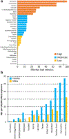
A) Lower extremity effective load ratings for common physical activities, with higher load ratings being representative of a greater bone osteogenic stimulus. Effective load ratings were estimated from the magnitude and rate of ground reaction force generation during representative actions (or similar actions when reaction forces could not be directly measured). Data from Weeks and Beck.[86] B) Incidence of bone stress injuries (BSIs) in females and males across collegiate sports in the United States over a 10-year period. N/A = data not available. Data from Rizzone et al.[15]
Figure 3.
Figure 3.
Loading-induced addition of bone on the outer periosteal surface is functionally important, helping the skeleton meet its dual needs of being strong to resist injury, but lightweight for energy efficient motion. A) The polar moment of inertia (i.e. strength) of a bone is proportional to the radii of its outer periosteal (rp) and inner endocortical (re) surfaces according to the relationship π(rp4-re4)/2. This relationship illustrates that periosteal surface changes have a greater influence on strength than changes on the endocortical surface. B) For example, a 5% increase in rp (equating to a 15% increase in bone mineral content [i.e. mass]) results in a disproportionate 24% increase in strength, assuming constant bone material properties (i.e. volumetric bone mineral density) and an initial rp-to-re ratio of 1.8. C) If the same mass of bone added to the periosteal surface was simultaneously removed from the endocortical surface, re would increase by 15%, but the bone would still be 16% stronger than the bone with same mass in A) because of its greater size (i.e. 5% greater rp). Broken lines in B) and C) indicate the original bone surfaces in A).
Figure 4.
Figure 4.
Vertical ground reaction force, and computed muscle generated and tibial compression forces during running with a typical rear-foot strike pattern. The external ground reaction force has two peaks—an initial rapidly reached impact peak (IP) and a second slower, but higher magnitude active peak (AP). Internal muscle generated and tibial compressive forces, computed via subject-specific musculoskeletal modeling, far exceed ground reaction forces and peak near the active peak of the ground reaction force. The later peak of tibial forces has raised the question of the relative contribution of initial foot-ground impact versus later muscle generated forces in BSI genesis. Image adapted from: Matijevich E, Scott L, Volgyesi P, Derry K, Zelik K. Combining wearable sensor signals, machine learning and biomechanics to estimate tibial bone force and damage during running. Human Movement Science 2020;74:102690, with permission from Elsevier.

Similar articles

Cited by

References

    1. Burr DB, Forwood MR, Fyhrie DP, Martin RB, Schaffler MB, Turner CH. Bone microdamage and skeletal fragility in osteoporotic and stress fractures. J Bone Miner Res. 1997;12:6–15. - PubMed
    1. Warden SJ, Davis IS, Fredericson M. Management and prevention of bone stress injuries in long-distance runners. J Orthop Sports Phys Ther. 2014;44(10):749–65. doi:10.2519/jospt.2014.5334. - DOI - PubMed
    1. Burr DB. Targeted and nontargeted remodeling. Bone. 2002;30(1):2–4. - PubMed
    1. Eriksen EF. Cellular mechanisms of bone remodeling. Rev Endocr Metab Disord. 2010;11(4):219–27. doi:10.1007/s11154-010-9153-1. - DOI - PMC - PubMed
    1. Nattiv A, Kennedy G, Barrack MT, Abdelkerim A, Goolsby MA, Arends JC et al. Correlation of MRI grading of bone stress injuries with clinical risk factors and return to play: a 5-year prospective study in collegiate track and field athletes. Am J Sports Med. 2013;41(8):1930–41. doi:10.1177/0363546513490645. - DOI - PMC - PubMed

Publication types