. 2021 Feb 26;17(2):e1009306.
doi: 10.1371/journal.pgen.1009306. eCollection 2021 Feb.

Defects in the cytoplasmic assembly of axonemal dynein arms cause morphological abnormalities and dysmotility in sperm cells leading to male infertility

Affiliations

Defects in the cytoplasmic assembly of axonemal dynein arms cause morphological abnormalities and dysmotility in sperm cells leading to male infertility

Isabella Aprea et al. PLoS Genet. .

Abstract

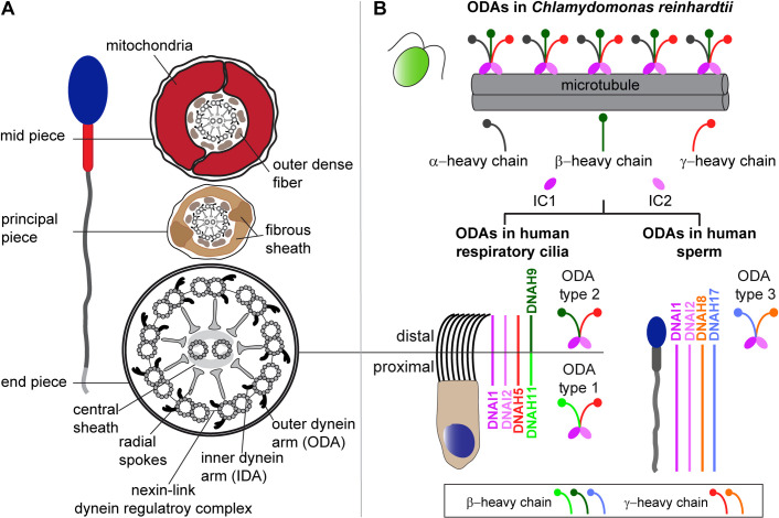
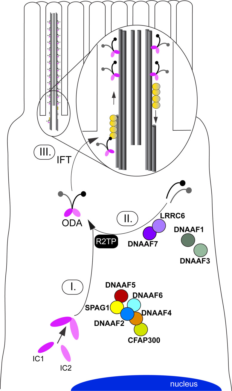
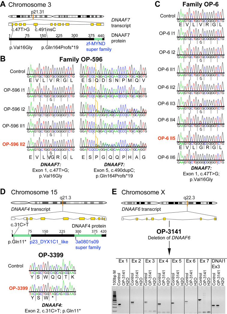
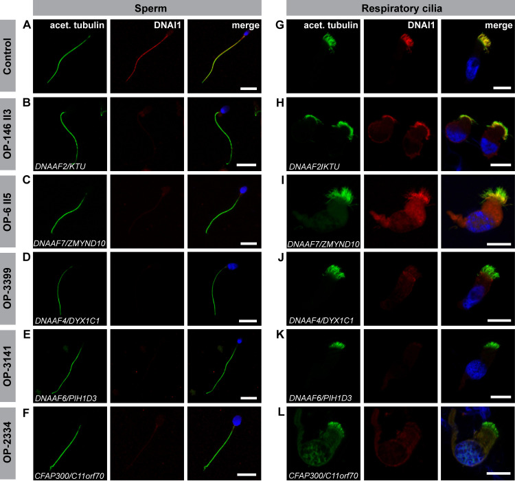
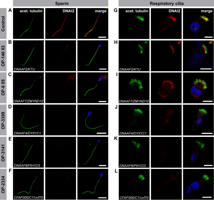
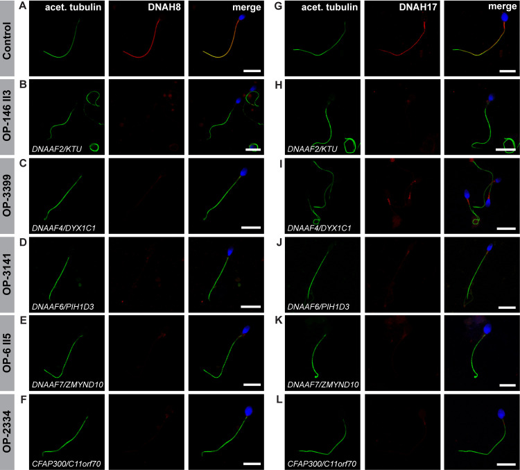
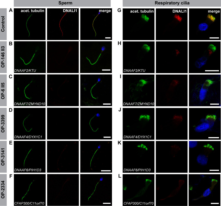
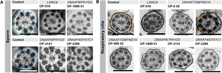
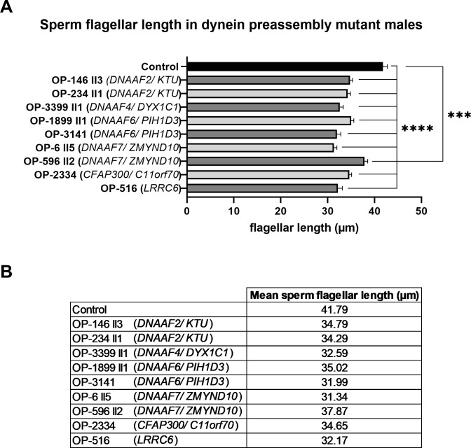
Axonemal protein complexes, such as outer (ODA) and inner (IDA) dynein arms, are responsible for the generation and regulation of flagellar and ciliary beating. Studies in various ciliated model organisms have shown that axonemal dynein arms are first assembled in the cell cytoplasm and then delivered into axonemes during ciliogenesis. In humans, mutations in genes encoding for factors involved in this process cause structural and functional defects of motile cilia in various organs such as the airways and result in the hereditary disorder primary ciliary dyskinesia (PCD). Despite extensive knowledge about the cytoplasmic assembly of axonemal dynein arms in respiratory cilia, this process is still poorly understood in sperm flagella. To better define its clinical relevance on sperm structure and function, and thus male fertility, further investigations are required. Here we report the fertility status in different axonemal dynein preassembly mutant males (DNAAF2/ KTU, DNAAF4/ DYX1C1, DNAAF6/ PIH1D3, DNAAF7/ZMYND10, CFAP300/C11orf70 and LRRC6). Besides andrological examinations, we functionally and structurally analyzed sperm flagella of affected individuals by high-speed video- and transmission electron microscopy as well as systematically compared the composition of dynein arms in sperm flagella and respiratory cilia by immunofluorescence microscopy. Furthermore, we analyzed the flagellar length in dynein preassembly mutant sperm. We found that the process of axonemal dynein preassembly is also critical in sperm, by identifying defects of ODAs and IDAs in dysmotile sperm of these individuals. Interestingly, these mutant sperm consistently show a complete loss of ODAs, while some respiratory cilia from the same individual can retain ODAs in the proximal ciliary compartment. This agrees with reports of solely one distinct ODA type in sperm, compared to two different ODA types in proximal and distal respiratory ciliary axonemes. Consistent with observations in model organisms, we also determined a significant reduction of sperm flagellar length in these individuals. These findings are relevant to subsequent studies on the function and composition of sperm flagella in PCD patients and non-syndromic infertile males. Our study contributes to a better understanding of the fertility status in PCD-affected males and should help guide genetic and andrological counselling for affected males and their families.


Conflict of interest statement

The authors have declared that no competing interests exist.

Figures

Fig 1
Fig 1. Structure of sperm flagella and respiratory cilia and ODA composition in both cell types.
(A) Cross section schemes of a sperm flagellum and multiciliated respiratory epithelial cell. Both cell types carry a microtubule-based 9+2 axoneme and multiprotein complexes serving as functional modules. Nine outer microtubule doublets surround the central pair and are connected by the nexin-link dynein regulatory complex. Radial spokes form the connecting bridge to the central pair and its associated protein complexes, the central sheath. Each outer doublet carries outer (ODA) and inner (IDA) dynein arms, important for flagellar and ciliary beat generation and regulation. Sperm flagella carry accessory structures that define specific flagellar regions. The mid piece contains a mitochondrial sheath and outer dense fibers surrounding the axoneme. In the sperm flagellar principal piece, the mitochondrial sheath is substituted by a fibrous sheath. In the flagellar end piece, the axoneme is surrounded solely by the plasma membrane. (B) ODAs of the green algae Chlamydomonas reinhardtii present a three-headed structure with an α-, β-, and γ-heavy chain (HC). Mammals present different ODA types, all characterized by a two-headed structure. Respiratory cilia carry ODA type 1 in the proximal and ODA type 2 in the distal ciliary compartment. In comparison to Chlamydomonas, both mammalian ODA types carry the γ-HC ortholog DNAH5, while the β-HC orthologs DNAH11 and DNAH9 are specific to type 1 and 2, respectively. ODA type 3 is specific to sperm flagella and carries DNAH8 as γ-HC and DNAH17 as β-HC ortholog. DNAI1 and DNAI2 are orthologs of Chlamydomonas intermediate chains (IC1 and 2) and are present in all three ODA types [17,18].
Fig 2
Fig 2. Schematic overview of the cytoplasmic dynein arm preassembly process in multiple motile cilia that is aided by distinct dynein axonemal assembly factors (DNAAFs).
Motile cilia protrude from the cell surface and contain microtubule-based axonemes. During ciliogenesis, axonemal components are pre-assembled in the cytoplasm (e.g. axonemal motor protein complexes, I.+II.) and subsequently delivered to the growing axonemes (III.) by intraflagellar transport (IFT) machinery. The figure representatively shows the interactions between DNAAFs (indicated in bold) as well as distinct outer dynein arm (ODA) components, as experimentally determined in previous studies [17,29,48,50,56]. HC: Heavy chains; IC: Intermediate chains.
Fig 3
Fig 3. Genetic testing in families OP-6, OP-596 and individuals OP-3141 and OP-3399 identifies disease causing mutations in the genes DNAAF7, DNAAF4 and DNAAF6, encoding axonemal dynein assembly factors.
(A) DNAAF7 (CCDS2825.1) is located on chromosome 3p21.31 (orange mark in the chromosome schematic) and consists of 12 exons encoding a 440 amino acid protein. A testis specific isoform comprising only 11 exons, due to selection of an alternative splicing acceptor site is annotated (CCDS77747.1). (B) Sanger sequencing of PCD family OP-596 confirms DNAAF7 compound heterozygous missense and insertion mutations (Exon 1: c.47T>G; p.Val16Gly and Exon 5: c.490dupC; p.Gln164Profs*19) in affected individual OP-596 II2 (indicated in dark orange). (C) Sanger sequencing of PCD family OP-6 confirms a DNAAF7 homozygous missense mutation (Exon 1: c.47T>G; p.Val16Gly) in affected individual OP-6 II5 (indicated in dark orange) and affected sibling OP-6 II6. (D) DNAAF4 (CCDS10154) is located on chromosome 15q21.3 (orange mark in the chromosome schematic) and consists of one non-coding and nine coding exons, encoding a 420 amino acid protein. Sanger sequencing (bottom of the figure) for PCD affected individual OP-3399 confirms a homozygous nonsense mutation in Exon 2 (c.31C>T; p.Gln11*). (E) DNAAF6 (CCDS14528) is located on the chromosome Xq22.3 (orange mark in the chromosome schematic) and consists of two non-coding and six coding exons, encoding a 214 amino acid protein. PCR amplification (lower figure in E) confirms a deletion of all DNAAF6 exons in PCD affected individual OP-3141 compared to control. To demonstrate DNA integrity of OP-3141 we amplified Exon 3 from the DNAI1 (ic: internal control) gene in OP-3141 and the control. Ex: exon; ins: insertion.
Fig 4
Fig 4. Dynein preassembly mutant sperm flagella and respiratory cilia show absence or severe reduction of the ODA intermediate chain DNAI1.
Control (A) and mutant (B-F) sperm cells, as well as control (G) and mutant (H-L) respiratory cells were double-labeled with antibodies directed against acetylated α tubulin (green) and the outer dynein arm intermediate chain DNAI1 (red). Both antibodies co-localize along the flagella and cilia in cells from the healthy control (yellow, A, G). In all mutant sperm cells, DNAI1 is not detected along the flagellar axoneme (B-F). In mutant respiratory cells of OP-146 II3 (H) and OP-6 II5 (I), DNAI1 is retained in the proximal ciliary length, whereas in mutant cells of OP-3399 (J), OP-3141 (K) and OP-2334 (L) DNAI1 displayed no immunoreactivity along the ciliary axoneme. Nuclei are stained with Hoechst33342 (blue). Scale bars represent 10 μm.
Fig 5
Fig 5. Dynein preassembly mutant sperm flagella and respiratory cilia show absence or reduction of the ODA intermediate chain DNAI2.
Control (A) and mutant (B-F) sperm, as well as control (G) and mutant (H-L) respiratory cells are double-labeled with antibodies directed against acetylated α tubulin (green) and the outer dynein arm intermediate chain DNAI2 (red). Both antibodies co-localize along the flagella and cilia in cells from the unaffected control (yellow, A, G). In all mutant sperm cells no signal for DNAI2 is observed in flagellar axonemes (B-F). In mutant respiratory cells of OP-146 II3 (H) and OP-6 II5 (I), DNAI2 localizes to the proximal part of the ciliary axoneme, whereas in mutant cells of OP-3399 (J), OP-3141 (K) and OP-2334 (L) immunoreactivity to DNAI2 is not observed in the ciliary axoneme. Nuclei are stained with Hoechst33342 (blue). Scale bars represent 10 μm.
Fig 6
Fig 6. Dynein preassembly mutant sperm flagella show absence or severe reduction of the sperm specific ODA heavy chains DNAH8 and DNAH17.
Control (A and G) and mutant sperm (B-F and H-L), deficient for the dynein preassembly factors DNAAF2, DNAAF4, DNAAF6, DNAAF7 and CFAP300, were double-labeled with antibodies directed against acetylated α tubulin (green), as flagellar marker, and the ODA heavy chains DNAH8 and DNAH17 (red). Both antibodies co-localize along the flagella from the unaffected control (merged image, A, G). In all mutant sperm flagella DNAH8 is absent from the flagellar axoneme (B-F). DNAH17 is either absent from the flagellar axoneme of mutant sperm (H, K, L) or severely reduced, displaying a residual signal in the flagellar midpiece (I, J), as observed in DNAAF4- and DNAAF6-deficient sperm. Nuclei are stained with Hoechst33342 (blue). Scale bars represent 10 μm.
Fig 7
Fig 7. Dynein preassembly mutant sperm flagella and respiratory cilia show absence of the IDA intermediate light chain DNALI1.
Control (A) and mutant (B-F) sperm, as well as control (G) and mutant (H-L) respiratory cells were double-labeled with antibodies directed against acetylated α tubulin (green) and the inner dynein arm intermediate light chain DNALI1 (red). Both antibodies co-localize along the flagella and cilia in cells from the unaffected control (yellow, A, G). In all mutant sperm flagella (B-F) and respiratory cells (H-L) DNALI1 is absent or severely reduced from the flagellar and ciliary axonemes. Nuclei are stained with Hoechst33342 (blue). Scale bars represent 10 μm.
Fig 8
Fig 8. Transmission electron microscopy (TEM) analysis confirms outer dynein arm (ODA) defects in sperm flagella and respiratory cilia from dynein preassembly mutant individuals.
(A) Electron micrographs of healthy control sperm show a regular flagellar 9+2 axoneme with clearly visible ODAs (indicated by blue arrowheads). Cross sections of sperm flagella of PCD-affected individuals (data shown for OP-516, OP-1899 II1, OP-3141 and OP-3399) confirm the absence and thus the ODA defect identified by high-resolution IF microscopy. (B) Analogously, axonemes of respiratory cilia of healthy control individuals show clearly visible and regularly disposed ODAs (indicated by orange arrowheads), whereas those of affected individuals lack the ODA complexes at their outer microtubule doublets. DNAAF7-mutant individual OP-6 II5 also displays some ciliary cross sections with ODAs still attached to microtubule doublets. Scale bars represent 200 nm.
Fig 9
Fig 9. The length of sperm flagella is reduced in dynein preassembly mutant sperm cells.
(A) Compared to control, sperm flagellar length is significantly reduced in DNAAF2- (OP-146 II3 and OP-234 II1), DNAAF4- (OP-3399), DNAAF6- (OP-1899 II1 and OP-3141), DNAAF7- (OP-6 II5 and OP-596 II2), CFAP300- (OP-2334) and LRRC6- (OP-516) mutant individuals. Statistical analysis was performed using a Brown-Forsythe ANOVA test on the entire dataset (p-value <0.0001: ****) and a two tailed t-test with Welch’s correction comparing each mutant individual to the control group (OP-596 II2 versus control: p-value 0.0009 (***); other mutant individuals versus control: p-value <0.0001 (****)). (B) Table with average value of sperm flagellar length for control sperm and the dynein preassembly mutant individuals analyzed. Error bars represent standard error mean (SEM).

References

    1. Tüttelmann F, Ruckert C, Röpke A. Spermatogenesestörungen: Perspektiven für erweiterte genetische Diagnostik nach 20 Jahren unveränderter Routine. Medizinische Genet. 2018;30: 12–20.
    1. WHO | Infertility is a global public health issue. WHO. 2019 [cited 4 Oct 2019]. Available from: https://www.who.int/reproductivehealth/topics/infertility/perspective/en/.
    1. Bracke A, Peeters K, Punjabi U, Hoogewijs D, Dewilde S. A search for molecular mechanisms underlying male idiopathic infertility. Reproductive BioMedicine Online. 2018;36(3): 327–339. 10.1016/j.rbmo.2017.12.005 - DOI - PubMed
    1. Kliesch S. Diagnosis of male infertility: Diagnostic work-up of the infertile man. European Urology, Supplements. 2014;13(4): 73–82.
    1. Krausz C. Male infertility: Pathogenesis and clinical diagnosis. Best Pract Res Clin Endocrinol Metab. 2011;25: 271–285. 10.1016/j.beem.2010.08.006 - DOI - PubMed

Publication types

MeSH terms