

Audiovisual integration in macaque face patch neurons
- PMID: 33636119
- PMCID: PMC8521527
- DOI: 10.1016/j.cub.2021.01.102
Audiovisual integration in macaque face patch neurons
Abstract
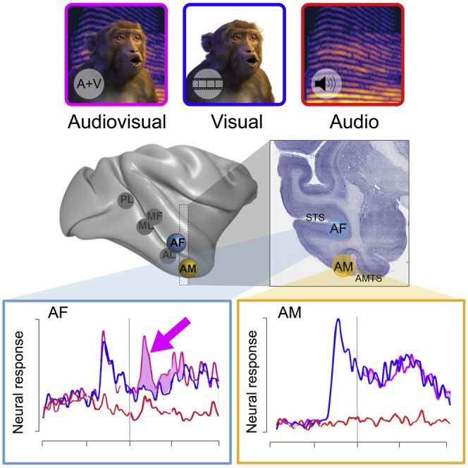
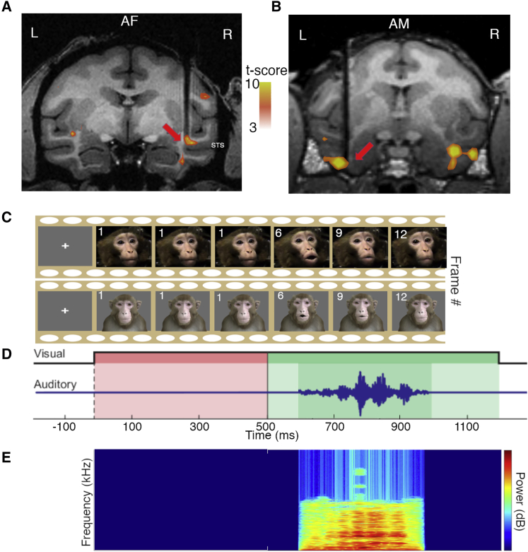
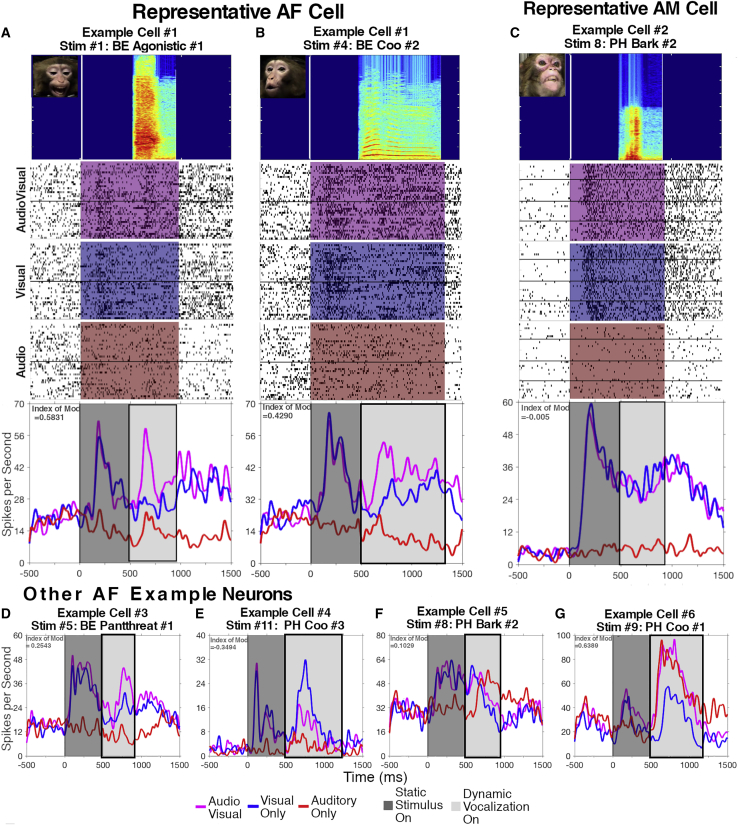
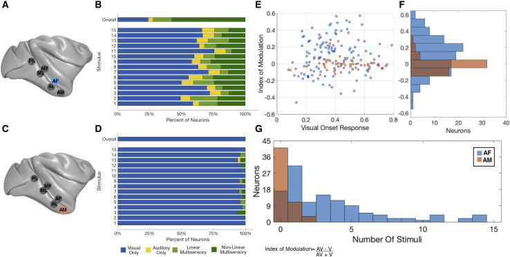
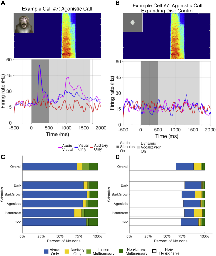
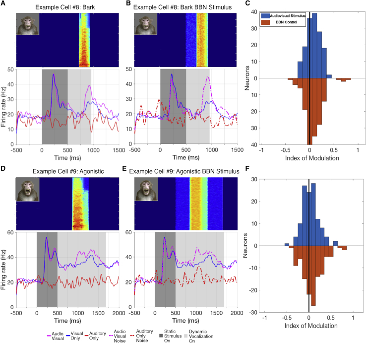
Primate social communication depends on the perceptual integration of visual and auditory cues, reflected in the multimodal mixing of sensory signals in certain cortical areas. The macaque cortical face patch network, identified through visual, face-selective responses measured with fMRI, is assumed to contribute to visual social interactions. However, whether face patch neurons are also influenced by acoustic information, such as the auditory component of a natural vocalization, remains unknown. Here, we recorded single-unit activity in the anterior fundus (AF) face patch, in the superior temporal sulcus, and anterior medial (AM) face patch, on the undersurface of the temporal lobe, in macaques presented with audiovisual, visual-only, and auditory-only renditions of natural movies of macaques vocalizing. The results revealed that 76% of neurons in face patch AF were significantly influenced by the auditory component of the movie, most often through enhancement of visual responses but sometimes in response to the auditory stimulus alone. By contrast, few neurons in face patch AM exhibited significant auditory responses or modulation. Control experiments in AF used an animated macaque avatar to demonstrate, first, that the structural elements of the face were often essential for audiovisual modulation and, second, that the temporal modulation of the acoustic stimulus was more important than its frequency spectrum. Together, these results identify a striking contrast between two face patches and specifically identify AF as playing a potential role in the integration of audiovisual cues during natural modes of social communication.
Keywords: audition; electrophysiology; face patches; multisensory integration; primate; vision.
Published by Elsevier Inc.
Conflict of interest statement
Declaration of interests The authors declare no competing interests.
Figures
Comment in
-
Face and voice perception: Monkey see, monkey hear.Curr Biol. 2021 May 10;31(9):R435-R437. doi: 10.1016/j.cub.2021.02.060. Curr Biol. 2021. PMID: 33974868
Similar articles
-
Auditory and visual modulation of temporal lobe neurons in voice-sensitive and association cortices.J Neurosci. 2014 Feb 12;34(7):2524-37. doi: 10.1523/JNEUROSCI.2805-13.2014. J Neurosci. 2014. PMID: 24523543 Free PMC article.
-
Timing of audiovisual inputs to the prefrontal cortex and multisensory integration.Neuroscience. 2012 Jul 12;214:36-48. doi: 10.1016/j.neuroscience.2012.03.025. Epub 2012 Apr 16. Neuroscience. 2012. PMID: 22516006 Free PMC article.
-
Dynamic Suppression of Average Facial Structure Shapes Neural Tuning in Three Macaque Face Patches.Curr Biol. 2021 Jan 11;31(1):1-12.e5. doi: 10.1016/j.cub.2020.09.070. Epub 2020 Oct 15. Curr Biol. 2021. PMID: 33065012 Free PMC article.
-
Multisensory interactions of face and vocal information during perception and memory in ventrolateral prefrontal cortex.Philos Trans R Soc Lond B Biol Sci. 2023 Sep 25;378(1886):20220343. doi: 10.1098/rstb.2022.0343. Epub 2023 Aug 7. Philos Trans R Soc Lond B Biol Sci. 2023. PMID: 37545305 Free PMC article. Review.
-
Coding of vocalizations by single neurons in ventrolateral prefrontal cortex.Hear Res. 2013 Nov;305:135-43. doi: 10.1016/j.heares.2013.07.011. Epub 2013 Jul 26. Hear Res. 2013. PMID: 23895874 Free PMC article. Review.
Cited by
-
Multisensory perception constrains the formation of object categories: a review of evidence from sensory-driven and predictive processes on categorical decisions.Philos Trans R Soc Lond B Biol Sci. 2023 Sep 25;378(1886):20220342. doi: 10.1098/rstb.2022.0342. Epub 2023 Aug 7. Philos Trans R Soc Lond B Biol Sci. 2023. PMID: 37545304 Free PMC article. Review.
-
Multilevel rhythms in multimodal communication.Philos Trans R Soc Lond B Biol Sci. 2021 Oct 11;376(1835):20200334. doi: 10.1098/rstb.2020.0334. Epub 2021 Aug 23. Philos Trans R Soc Lond B Biol Sci. 2021. PMID: 34420378 Free PMC article. Review.
-
Socially meaningful visual context either enhances or inhibits vocalisation processing in the macaque brain.Nat Commun. 2022 Aug 19;13(1):4886. doi: 10.1038/s41467-022-32512-9. Nat Commun. 2022. PMID: 35985995 Free PMC article.
-
Representation of Expression and Identity by Ventral Prefrontal Neurons.Neuroscience. 2022 Aug 1;496:243-260. doi: 10.1016/j.neuroscience.2022.05.033. Epub 2022 May 30. Neuroscience. 2022. PMID: 35654293 Free PMC article.
-
Mapping of facial and vocal processing in common marmosets with ultra-high field fMRI.Commun Biol. 2024 Mar 13;7(1):317. doi: 10.1038/s42003-024-06002-1. Commun Biol. 2024. PMID: 38480875 Free PMC article.
References
-
- Ghazanfar A.A., Santos L.R. Primate brains in the wild: the sensory bases for social interactions. Nat. Rev. Neurosci. 2004;5:603–616. - PubMed
-
- Beauchamp M.S., Lee K.E., Argall B.D., Martin A. Integration of auditory and visual information about objects in superior temporal sulcus. Neuron. 2004;41:809–823. - PubMed
-
- Beauchamp M.S., Argall B.D., Bodurka J., Duyn J.H., Martin A. Unraveling multisensory integration: patchy organization within human STS multisensory cortex. Nat. Neurosci. 2004;7:1190–1192. - PubMed
-
- Barraclough N.E., Xiao D., Baker C.I., Oram M.W., Perrett D.I. Integration of visual and auditory information by superior temporal sulcus neurons responsive to the sight of actions. J. Cogn. Neurosci. 2005;17:377–391. - PubMed
Publication types
MeSH terms
Grants and funding
LinkOut - more resources
Full Text Sources
Other Literature Sources
