. 2021 Mar 4;184(5):1262-1280.e22.
doi: 10.1016/j.cell.2021.02.019. Epub 2021 Feb 25.

In vivo CD8+ T cell CRISPR screening reveals control by Fli1 in infection and cancer

Affiliations

In vivo CD8+ T cell CRISPR screening reveals control by Fli1 in infection and cancer

Zeyu Chen et al. Cell. .

Abstract

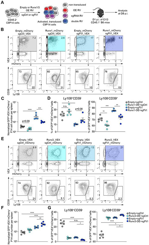
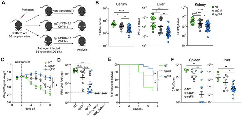
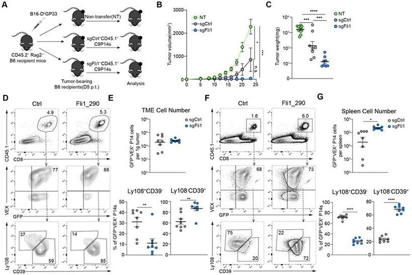
Improving effector activity of antigen-specific T cells is a major goal in cancer immunotherapy. Despite the identification of several effector T cell (TEFF)-driving transcription factors (TFs), the transcriptional coordination of TEFF biology remains poorly understood. We developed an in vivo T cell CRISPR screening platform and identified a key mechanism restraining TEFF biology through the ETS family TF, Fli1. Genetic deletion of Fli1 enhanced TEFF responses without compromising memory or exhaustion precursors. Fli1 restrained TEFF lineage differentiation by binding to cis-regulatory elements of effector-associated genes. Loss of Fli1 increased chromatin accessibility at ETS:RUNX motifs, allowing more efficient Runx3-driven TEFF biology. CD8+ T cells lacking Fli1 provided substantially better protection against multiple infections and tumors. These data indicate that Fli1 safeguards the developing CD8+ T cell transcriptional landscape from excessive ETS:RUNX-driven TEFF cell differentiation. Moreover, genetic deletion of Fli1 improves TEFF differentiation and protective immunity in infections and cancer.

Keywords: CD8; CRISPR; Fli1; cancer; chronic infection; effector CD8 T cell; exhausted CD8 T cell; exhaustion; immunotherapy; protective immunity.


Conflict of interest statement

Declaration of interests E.J.W. has consulting agreements with and/or is on the scientific advisory board for Merck, Elstar, Janssen, Related Sciences, Synthekine and Surface Oncology. E.J.W. is a founder of Surface Oncology and Arsenal Biosciences. E.J.W. has a patent licensing agreement on the PD-1 pathway with Roche/Genentech. O.K. is an employee and shareholder of Arsenal Biosciences.

Figures

Figure 1.
Figure 1.. Dissecting transcriptional programs of CD8+ T cells using the OpTICS system.
A. Experimental design for Optimized T cell In vivo CRISPR Screening (OpTICS). On Day 0(D0), CD8+ T cells were isolated from CD45.2+ C9P14 mice and activated in vitro; CD45.1+ WT recipient mice were infected with LCMV. On D1 p.i., activated C9P14 cells were transduced with the RV-sgRNA library for 6 hours. On D2 p.i., Cas9+sgRNA+ P14 cells were purified, 5-10% of the sorted cells frozen for D2 baseline (T0 time point), and the rest were adoptively transferred into LCMV-infected recipient mice. Cas9+sgRNA+ P14 cells were isolated from recipient mice on the indicated days by MACS and FACS (T1 time point). Targeted PCR with sequencing adaptors for the sgRNA cassettes was performed and PCR products were sequenced. The CRISPR Score (CS) was calculated as shown. B. The CS comparing T1 time point to T0 time point (D2 baseline) for target genes from Cas9+sgRNA+ cells from spleen on D8 or D15 p.i. of Arm, D9 or D14 p.i. of Cl13. X-axis shows targeted genes; y-axis shows the CS of each targeted gene (using 4-5 sgRNAs). C. Heatmap of CS for targeted genes. Heatmap ranks the geometric means of CS for each gene. See Star Methods. D. Distribution of Ctrl, Pdcd1 and Fli1 sgRNAs. Axis represents log2 fold change (FC). Histogram shows distribution of all sgRNAs. Red bars represent targeted sgRNAs, grey bars represent all other sgRNAs. E. Western blot for Fli1 protein from sorted Cas9+sgFli1+ P14 cells and paired Cas9+sgFli1 P14 cells from spleen. Two Fli1-sgRNAs (sgFli1_290 and sgFli1_360) were used. Pooled mice were used (3-5 mice for Arm, 10-15 mice for Cl13). Bar graph represents the normalized band intensity of Fli1. Fli1 was first normalized to GAPDH, then a ratio between Cas9+sgFli1+ versus Cas9+sgFli1 was calculated and is shown. F. Normalized Cas9+sgRNA (VEX)+ cell numbers from spleen for Ctrl-sgRNA(sgCtrl) and 2 Fli1-sgRNA (sgFli1_290 and sgFli1_360) groups on D8 p.i. of Arm, D15 p.i. of Arm, D9 p.i. of Cl13 and D14 p.i. of Cl13. Cell numbers normalized to the sgCtrl group based on D2 in vitro transduction efficiency (see Figure S3B and S3D). *P<0.05, **P<0.01, ***P<0.001, ****P<0.001 versus control (One-Way Anova analysis). Data are representative of 4 independent experiments (mean±s.e.m.) with at least 4 mice/group for F.
Figure 2.
Figure 2.. Fli1 restrains TEFF cell proliferation and differentiation during acute infection.
A. Flow cytometry plots and statistical analysis of KLRG1HiCD127Lo Terminal Effectors (TEs) and KLRG1LoCD127Hi Memory Precursors (MPs). Frequencies (left) and numbers (right) from spleen for sgCtrl and 2 sgFli1 groups on D8 and D15 p.i. of Arm. Gated on Cas9(GFP)+ sgRNA(VEX)+ P14 cells. B. Flow cytometry plots and statistical analysis of CX3CR1+CXCR3 TEFF cells and CX3CR1CXCR3+ early TMEM cell frequencies (left) and numbers (right) from spleen for sgCtrl and 2 sgFli1 groups on D8 and D15 p.i. of Arm. Gated on Cas9+sgRNA+ P14 cells. (C-E) Experimental design. On D0, CD45.1+ P14 cells were activated and recipient mice were infected with Arm; On D1 p.i., activated P14 cells were transduced with Empty-RV or Fli1-over expressing (OE)-RV. On D2 p.i., VEX+ P14 cells were purified for each group, and 5 x 10⁴ cells were adoptively transferred into infected recipient mice. C. Flow cytometry plots of CD45.2+VEX+ cell frequency and statistical analysis of CD45.2+VEX+ cell number for Empty-RV and Fli1-OE-RV conditions. D. Flow cytometry plots and statistical analysis of KLRG1HiCD127Lo TE and KLRG1LoCD127Hi MP frequencies from spleen for Empty-RV and Fli1-OE-RV groups on D8 and D15 p.i. of Arm. Gated on VEX+ P14 cells. E. Flow cytometry plots and statistical analysis of CX3CR1+CXCR3 TEFF cell and CX3CR1CXCR3+ early TMEM cell frequencies from spleen for Empty-RV and Fli1-OE-RV groups on D8 and D15 p.i. of Arm. Gated on VEX+ P14 cells. *P<0.05, **P<0.01, ***P<0.001, ****P<0.001 versus control (two-tailed Student’s t-test and One-Way Anova analysis). Data are representative of 2-4 independent experiments (mean±s.e.m.) with at least 3 mice/group.
Figure 3.
Figure 3.. Fli1 antagonizes TEFF-like cell differentiation during chronic infection.
A. Flow cytometry plots and statistical analysis of Ly108CD39+ or TCF-1Gzmb+ TEFF-like cells and Ly108+CD39 or TCF-1+Gzmb TEX precursor frequencies from spleen for sgCtrl and 2 sgFli1 groups on D8 and D15 p.i. of Cl13. Gated on the Cas9(GFP)+sgRNA(VEX)+ P14 cells. B. Statistical analysis of CX3CR1+ and Tim-3+ frequencies, and KLRG1 and PD-1 MFI from spleen for sgCtrl and 2 sgFli1 groups on D8 and D15 p.i. of Cl13. Gated on the Cas9+sgRNA+ P14 cells. C. Heatmap of differentially expressed genes between sgCtrl and 2 sgFli1 groups. D. Gene Ontology (GO) enrichment analysis for the sgFli1 groups. E. GO enrichment analysis for the sgCtrl groups. F. Gene Set Enrichment Analysis (GSEA) of TEX precursor signature (adapted from Chen et al. 2019) between sgCtrl and sgFli1 groups. G. GSEA of TEFF-like signature (adapted from Bengsch et al. 2018) between sgCtrl and sgFli1 groups. *P<0.05, **P<0.01, ***P<0.001, ****P<0.001 versus control (two-tailed Student’s t-test and One-Way Anova analysis). Data are representative of 4 independent experiments (mean±s.e.m.) with at least 4 mice/group for A and B.
Figure 4.
Figure 4.. Fli1 reshapes the epigenetic profile of CD8+ T cells and inhibits TEFF-associated gene expression.
A. PCA plot of ATAC-seq data for sgCtrl, sgFli1_290 and sgFli1_360 groups on D9 p.i. of Cl13. B. Overall open chromatin region (OCR) peak changes for sgFli1 groups compared to sgCtrl group. C. Categories of Cis-element OCR peaks that changed between sgCtrl and sgFli1 groups. Left plot represents all changes; right plot represents changes for increased or decreased accessibility. D. Heatmap shows differentially accessible peaks between sgCtrl group and 2 sgFli1 groups (adjusted p-value<0.05, Log10 Fold Change>0.6). Selected genes assigned to the peaks are indicated. E. Overlapping Venn plot of the genes with differentially accessible (DA) peaks and differentially expressed genes from Figure 3C. F. Pearson correlation of the peak accessibility of the nearest genes versus the differential expression of the genes. G. Transcription factor (TF) motif gain or loss associated with loss of Fli1. X-axis represents the logP-value of the motif enrichment. Y-axis represents the fold change of the motif enrichment. Targeted motifs in the changed OCR between the sgCtrl and sgFli1 groups were compared to the whole genome background to calculate p-value and fold change. H. IgG or Fli1 binding signals from CUT&RUN on P14 cells on D8 p.i. of Cl13 and OCR signals detected by ATAC-seq for sgCtrl-sgRNA, sgFli1_290 and sgFli1_360 groups at the Cd28, Cx3cr1, and Havcr2 loci. I. Histogram of CD28 staining and statistical analysis for sgCtrl, sgFli1_290 and sgFli1_360 groups on D8 Cl13 p.i. Grey shows CD28 staining of CD44 naïve T cell. J. Heatmap shown differentially accessible (DA) peaks overlapped with the Fli1 CUT&RUN binding peaks between sgCtrl group and 2 sgFli1 groups. Selected genes assigned to the peaks are indicated. K. Top 4 enriched TF motifs in the Fli1 CUT&RUN peaks are shown. *P<0.05, **P<0.01 versus control (One-Way Anova analysis). Data are representative of 2 independent experiments (mean±s.e.m.) with at least 5 mice/group for I.
Figure 5
Figure 5. Overexpression of Runx1 or Runx3 in the context of Fli1-deficiency in CD8+ T cells.
A. Experimental design. On D0, CD8+ T cells were isolated from CD45.2+ C9P14 donor mice and activated; CD45.1+ WT recipient mice were infected with Cl13. On D1 p.i., activated C9P14 cells were transduced with sgRNA-RV or OE-RV and 1 x 10⁵ transduced cells adoptively transferred into infected recipient mice. B-D. Flow cytometry plots (B) and statistical analysis (C-D) of VEX+mCherry+ C9P14 cells and Ly108CD39+/Ly108+CD39 C9P14 cells from spleen for sgCtrl-VEX + Empty-mCherry, sgCtrl-VEX + Runx1-mCherry, sgFli1_290-VEX + Empty-mCherry and sgFli1_290-VEX + Runx1-mCherry on D8 p.i. of Cl13. Gated on Cas9(GFP)+CD45.2+ P14 cells. E-G. Flow cytometry plots (E) and statistical analysis (F-G) of VEX+mCherry+ C9P14 cells and Ly108CD39+/Ly108+CD39 C9P14 cells from spleen for sgCtrl-mCherry + Empty-VEX, sgCtrl-mCherry + Runx3-VEX, sgFli1_290-mCherry + Empty-VEX and sgFli1_290-mCherry + Runx3-VEX at D8 p.i. of Cl13. Gated on Cas9+CD45.2+ P14 cells. *P<0.05, **P<0.01, ***P<0.001, ****P<0.001 versus control (One-Way Anova analysis). Data are representative of 2 independent experiments (mean±s.e.m.) with at least 5 mice/group.
Figure 6.
Figure 6.. Fli1-deficiency in CD8+ T cells enhances protective immunity against infections.
A. Experimental design. On D0, CD8+ T cells were isolated from CD45.2+ C9P14 donor mice and activated; CD45.1+ WT recipient mice were infected with LCMV Cl13, influenza virus PR8-GP33 or Listeria monocytogenes-GP33 (LM-GP33). On D1 p.i., activated C9P14 cells were transduced with sgCtrl or sgFli1 RV. On D2 p.i., Cas9+sgRNA(VEX)+ P14 cells were purified by flow cytometry for sgCtrl or sgFli1 groups, and adoptively transferred into the infected recipient mice. For Cl13, 1.5 x 10⁵ VEX+ C9P14 cells were transferred per mouse; for PR8-GP33 and LM-GP33, 1.0 x 10⁵ VEX+ C9P14 cells were transferred per mouse. B. LCMV viral load was measured by plaque assay on D15 p.i. with Cl13 in liver, kidney and serum of the indicated mice. Data pooled from 2 independent experiments. C. Weight curve for PR8-GP33 infected mice from NT, sgCtrl+ cell-transferred, or sgFli1+ cell-transferred groups. Dashed line represents the time of C9P14 adoptive transfer. D. PR8-GP33 viral RNA load in the lung of NT, sgCtrl+ or sgFli1+ C9P14 recipient mice. Dashed line indicates the limit of detection. Lung samples from naïve mice and spleen samples from PR8-GP33 infected mice were used as negative controls. E. Adjusted survival curve of LM-GP33 infected mice for NT, sgCtrl+ or sgFli1+ C9P14 recipient mice. Dashed line represents the time of C9P14 adoptive transfer. F. LM-GP33 bacteria load in spleen and liver of the surviving NT, sgCtrl+ or sgFli1+ C9P14 recipient mice on D7 p.i. *P<0.05, **P<0.01, ***P<0.001, ****P<0.001 versus control (One-Way Anova analysis for 6B-6D, 6F and Mantel-Cox test for 6E). Data are representative of 2 independent experiments (mean±s.e.m.) with at least 3 mice/group.
Figure 7.
Figure 7.. Loss of Fli1 in CD8+ T cells improved anti-tumor immunity.
A. Experimental design. On D0, CD45.2+Rag2−/− mice were inoculated with 1.0 x 10⁵ B16-Dbgp33 cells. On D3 post tumor inoculation (p.t.), CD8+ T cells were isolated from CD45.1+ C9P14 mice and activated. On D4 p.t., activated C9P14 cells were transduced with sgCtrl or sgFli1 RVs. On D5 p.t., sgRNA (VEX)+Cas9 (GFP)+P14 cells were sorted from sgCtrl or sgFli1 groups, and 1 x 10⁶ purified VEX+ C9P14 cells were adoptively transferred into tumor-bearing mice. B. Tumor volume curve for mice receiving NT, sgCtrl+ or sgFli1+ C9P14 cells. C. Tumor weight on D23 p.t. for mice receiving NT, sgCtrl+ or sgFli1+ C9P14 cells. D-E. Flow cytometry plots (D) and statistical analysis (E) of CD45.1+ sgRNA(VEX)+Cas9+P14 cells and Ly108CD39+/Ly108+CD39 C9P14 cells from tumor for sgCtrl or sgFli1 groups on D23 p.t. F-G. Flow cytometry plots (F) and statistical analysis (G) of CD45.1+ sgRNA+Cas9+ P14 cells and Ly108CD39+/Ly108+CD39 C9P14 cells from spleen for sgCtrl or sgFli1 groups at D23 p.t. *P<0.05, **P<0.01, ***P<0.001, ****P<0.001 versus control (two-tailed Student’s t-test and One-Way Anova analysis). Data are representative of 2 independent experiments (mean±s.e.m.) with at least 5 mice/group.

Comment in

References

    1. Alfei F, Kanev K, Hofmann M, Wu M, Ghoneim HE, Roelli P, Utzschneider DT, Hösslin, von M, Cullen JG, Fan Y, et al. (2019). TOX reinforces the phenotype and longevity of exhausted T cells in chronic viral infection. Nature 2017 545:7652 1. - PubMed
    1. Angelosanto JM, Blackburn SD, Crawford A, and Wherry EJ (2012). Progressive loss of memory T cell potential and commitment to exhaustion during chronic viral infection. J. Virol 86, 8161–8170. - PMC - PubMed
    1. Badovinac VP, Haring JS, and Harty JT (2007). Initial T Cell Receptor Transgenic Cell Precursor Frequency Dictates Critical Aspects of the CD8+ T Cell Response to Infection. Immunity 26, 827–841. - PMC - PubMed
    1. Badovinac VP, Porter BB, and Harty JT (2004). CD8+ T cell contraction is controlled by early inflammation. Nature Immunology 2006 7:12 5, 809–817. - PubMed
    1. Barber DL, Wherry EJ, Masopust D, Zhu B, Allison JP, Sharpe AH, Freeman GJ, and Ahmed R (2006). Restoring function in exhausted CD8 T cells during chronic viral infection. Nature 2017 545:7652 439, 682–687. - PubMed

Publication types

Substances