. 2021 Feb 26;27(1):20.
doi: 10.1186/s10020-021-00268-5.

Exosomal miR-335 derived from mature dendritic cells enhanced mesenchymal stem cell-mediated bone regeneration of bone defects in athymic rats

Affiliations

Exosomal miR-335 derived from mature dendritic cells enhanced mesenchymal stem cell-mediated bone regeneration of bone defects in athymic rats

Zhongliu Cao et al. Mol Med. .

Abstract

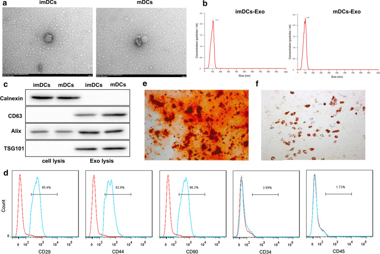
Background: Transplantation of bone marrow-derived mesenchymal stem cells (BM-MSCs) embedded in a bio-compatible matrix has been demonstrated as a promising strategy for the treatment of bone defects. This study was designed to explore the effect and mechanism of exosomes derived from mature dendritic cells (mDC-Exo) on the BM-MSCs-mediated bone regeneration using the matrix support in an athymic rat model of femoral bone defect.

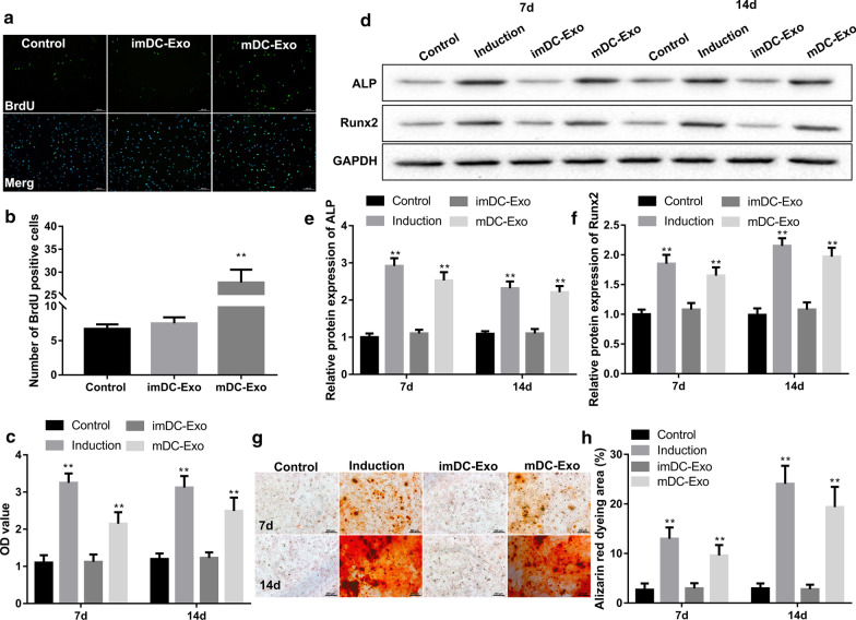
Methods: The BM-MSCs were isolated from rats and incubated with osteoblast induction medium, exosomes derived from immature DCs (imDC-Exo), mDC-Exo, and miR-335-deficient mDC-Exo. BM-MSCs treated without or with mDC-Exo were embedded in a bio-compatible matrix (Orthoss®) and then implanted into the femoral bone defect of athymic rats.

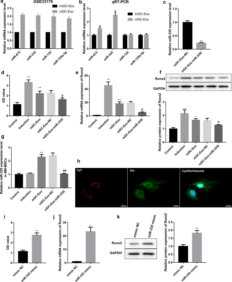
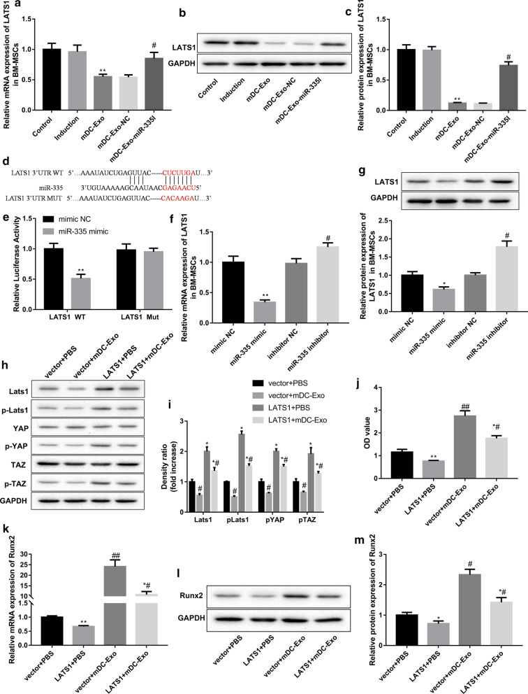
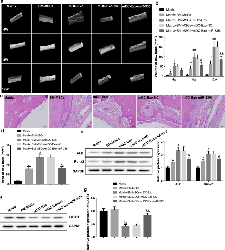
Results: mDC-Exo promoted the proliferation and osteogenic differentiation of BM-MSCs by transferring miR-335. Mechanistically, exosomal miR-335 inhibited Hippo signaling by targeting large tongue suppressor kinase 1 (LATS1) and thus promoted the proliferation and osteogenic differentiation of BM-MSCs. Animal experiments showed that mDC-Exo enhanced BM-MSCs-mediated bone regeneration after bone defect, and this effect was abrogated when miR-335 expression was inhibited in mDC-Exo.

Conclusion: mDC-Exo promoted osteogenic differentiation of BM-MSCs and enhanced BM-MSCs-mediated bone regeneration after femoral bone defect in athymic rats by transferring miR-335.

Keywords: Bone defect; Dendritic cell; Exosome; Mesenchymal stem cells; miR-335.


Conflict of interest statement

The authors declare that they have no competing interest.

Figures

Fig. 1
Fig. 1
Identification of exosomes and BM-MSCs. The exosomes isolated from imDCs and mDCs were referred to as imDCs-Exo and mDCs-Exo, respectively. a The morphological characterization of imDCs-Exo and mDCs-Exo by TEM. b The particle size of imDCs-Exo and mDCs-Exo measured using nanoparticle tracking analysis. c Expression of the exosomal markers (CD63, and Alix) and the exosome negative marker (Calnexin) confirmed by western blot. d The surface antigens including CD29, CD44, CD90, CD34, and CD45 of rat BM-MSCs was detected by flow cytometry. The positive rate of each surface marker was presented. e Osteogenic differentiation of BM-MSCs was confirmed by Alizarin Red staining. f Adipogenic differentiation of BM-MSCs was identified using Oil Red O staining
Fig. 2
Fig. 2
mDC-Exo promoted proliferation and osteogenic differentiation of BM-MSCs. a Immunofluorocytochemical staining for BrdU (green) and DNA (DAPI, blue) in BM-MSCs and b quantitation of the number of BrdU-positive cells in BM-MSCs in the groups of control, imDC-Exo, and mDC-Exo. c ALP activities expressed in optical density (OD) values at 520 nm, d–f protein levels of ALP and Runx2 determined by western blot, g, h representative Alizarin red staining assessing calcium deposits and quantification of the percentage of Alizarin red dyeing area in BM-MSCs in the groups of control, induction, imDC-Exo, and mDC-Exo on the 7th and 14th day of culture. **P < 0.01, vs. Control
Fig. 3
Fig. 3
mDC-Exo promoted osteogenic differentiation of BM-MSCs by transferring miR-335. a Higher expression of miR-672, miR-335, miR-124, and miR-125a-5p in mouse mDC-Exo relative to imDC-Exo (GSE33179). *P < 0.05, vs. imDC-Exo. b Expression of miR-672, miR-335, miR-124, and miR-125a-5p determined by qRT-PCR analysis in imDC-Exo and mDC-Exo separated according to the method mentioned above. *P < 0.05, vs. imDC-Exo. c miR-335 expression determined by qRT-PCR analysis in mDC-Exo-NC and mDC-Exo-miR-335I. **P < 0.01, vs. mDC-Exo-NC. d ALP activities expressed in optical density (OD) values at 520 nm, e, f Runx2 mRNA and protein levels, and g miR-335 expression in BM-MSCs in the groups of control, induction, mDC-Exo, and mDC-Exo-NC, and mDC-Exo-miR-335I on the 7th day of culture. *P < 0.05, **P < 0.01, vs. Control; #P < 0.05, ##P < 0.01, vs. mDC-Exo-NC. h The exosomes isolated from Cy3 (red)-miR-335-transfected mDCs were labeled with the lipophilic fluorescent dye Dio (green) and incubated with BM-MSCs (nuclei stained with Hoechst 33,342, blue). The uptake of exosomes by BM-MSCs was observed by confocal laser microscopy. (I) ALP activities expressed in OD values at 520 nm, (J-K) Runx2 mRNA and protein levels in BM-MSCs transfected with miR-335 mimics and mimic NC. **P < 0.01, vs. mimic NC
Fig. 4
Fig. 4
Exosomal miR-335 promoted osteogenic differentiation of BM-MSCs by targeting LATS1. LATS1 mRNA (a) and protein (b, c) levels in BM-MSCs in the groups of control, induction, mDC-Exo, and mDC-Exo-NC, and mDC-Exo-miR-335I on the 7th day of culture. **P < 0.01, vs. Control; #P < 0.05, vs. mDC-Exo-NC. d Predicted binding sites for miR-335 in the 3′-UTR of LATS1 (Targetscan). e The interaction between miR-335 and LAST1 3′-UTR was analyzed by luciferase activity assay. **P < 0.01, vs. mimic NC. LAST1 mRNA (f) and protein (g) levels in BM-MSCs transfected with miR-335 mimics, mimic NC, miR-335 inhibitors, or inhibitor NC. *P < 0.05, **P < 0.01, vs. mimic NC; #P < 0.05, vs. inhibitor NC. h, i The protein levels of LATS1, p-LAST1, YAP, p-YAP, TAZ, and p-TAZ, j ALP activities expressed in optical density (OD) values at 520 nm, k–m Runx2 mRNA and protein levels in BM-MSCs transfected with LAST1 overexpression vector/empty vector and treated with mDC-Exo or PBS. *P < 0.05, **P < 0.01, vs. vector + PBS; #P < 0.05, ##P < 0.01, vs. vector + mDC-Exo
Fig. 5
Fig. 5
mDC-Exo enhanced BM-MSCs-mediated bone regeneration by transferring miR-335. Rats were randomly divided into five groups: Matrix, Matrix + BM-MSCs, Matrix + BM-MSCs + mDC-Exo, Matrix + BM-MSCs + mDC-Exo-NC, and Matrix + BM-MSCs + mDC-Exo-miR-335I. a Bone regeneration was monitored via micro-CT scanning at 4, 8 and 12 weeks post-surgery. b Quantification of new bone volume from micro-CT scanning. c Representative H&E staining showing bone defect repair of posterior limbs of rats. d Quantification of new bone area from H&E staining. The protein levels of e ALP, Runx2, and f, g LAST1 in the bone examined by western blot. *P < 0.05, **P < 0.01, vs. Matrix; #P < 0.05, ##P < 0.01, vs. Matrix + BM-MSCs; $P < 0.05, $$P < 0.01, vs. Matrix + BM-MSCs + mDC-Exo-NC

Similar articles

Cited by

References

    1. Al-Moraissi EA, Oginni FO, Mahyoub Holkom MA, Mohamed AAS, Al-Sharani HM. Tissue-engineered bone using mesenchymal stem cells versus conventional bone grafts in the regeneration of maxillary alveolar bone: a systematic review and meta-analysis. Int J Oral Maxillofacial Implants. 2020;35(1):79–90. doi: 10.11607/jomi.7682. - DOI - PubMed
    1. An J, Li G, Zhang J, Zhou H, Jiang J, Wang X, Feng X, Wang S. GNAS knockdown suppresses osteogenic differentiation of mesenchymal stem cells via activation of Hippo signaling pathway. J Cell Physiol. 2019;234(12):22299–22310. doi: 10.1002/jcp.28796. - DOI - PubMed
    1. Burastero G, Scarfì S, Ferraris C, Fresia C, Sessarego N, Fruscione F, Monetti F, Scarfò F, Schupbach P, Podestà M, et al. The association of human mesenchymal stem cells with BMP-7 improves bone regeneration of critical-size segmental bone defects in athymic rats. Bone. 2010;47(1):117–126. doi: 10.1016/j.bone.2010.03.023. - DOI - PubMed
    1. Cañas JA, Sastre B, Rodrigo-Muñoz JM, Del Pozo V. Exosomes: A new approach to asthma pathology. Clin Chim Acta. 2019;495:139–147. doi: 10.1016/j.cca.2019.04.055. - DOI - PubMed
    1. Chen Z, Wu C, Gu W, Klein T, Crawford R, Xiao Y. Osteogenic differentiation of bone marrow MSCs by β-tricalcium phosphate stimulating macrophages via BMP2 signalling pathway. Biomaterials. 2014;35(5):1507–1518. doi: 10.1016/j.biomaterials.2013.11.014. - DOI - PubMed

Publication types

MeSH terms

LinkOut - more resources